Figures & data
Figure 1. Characterization of EPCs and EPC-MVs. (a) EPCs were identified by Di-LDL and Bs-Lectin staining. (b) Representative image of EPC-MVs and EPC-MVs-miR126 examined by TEM. (c) Size distribution of EPC-MVs and EPC-MVs-miR126 detected by NTA. (d) EPC-MVs stained with PE-CD34 and PE-VEGFR were analysed by flow cytometry.
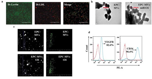
Figure 2. The expression of miR126 in different groups. **p< 0.01 vs. EPC, ##p< 0.01 vs. EPC-MVs.
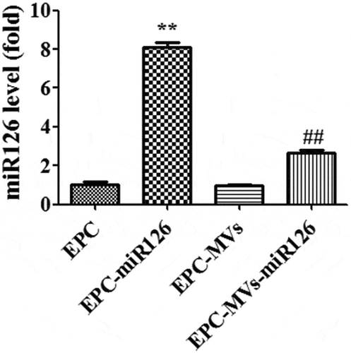
Figure 3. The effects of EPC-MVs and EPC-MVs-miR126 on the proliferation of MC3T3-E1 cells. (a) The effects of different concentrations of MVs on MC3T3-E1 cell viability at 48 h. (b) The proliferation of MC3T3-E1 cell in different groups. *p< 0.05, **p< 0.01 vs. control; #p< 0.05 vs. EPC-MVs; ++p< 0.01 vs. EPC-MVs-miR126; aap<0.01 vs. EPC-MVs; bp<0.05 vs. EPC-MVs+PD98059.
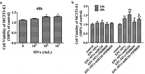
Figure 4. The effects of EPC-MVs and EPC-MVs-miR126 on the migration of MC3T3-E1 cells. (a) The expression of miR126 before and after wounding. (b) Representative image of cell migration at 48 h of MVs treatment. (c) Summarized data on cell migration. **p< 0.01 vs. control; ##p< 0.01 vs. EPC-MVs; ++p< 0.01 vs. EPC-MVs-miR126; aap<0.01 vs. EPC-MVs; bbp<0.01 vs. EPC-MVs+PD98059.
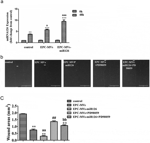
Figure 5. The effects of EPC-MVs and EPC-MVs-miR126 on apoptosis of MC3T3-E1 cell. (a) Cell apoptosis was measured by flow cytometry. (b) Cell apoptosis was determined by Hoechst 33258 staining (Red arrows represent apoptosis cells, white arrows represent normal cells). (c) and (d) Summarized data on apoptosis rate (%) assessed by flow cytometry and Hoechst 33258 staining. *p< 0.05, **p< 0.01 vs. SD; ##p< 0.01 vs. EPC-MVs; ++p< 0.01 vs. EPC-MVs-miR126; aap<0.01 vs. EPC-MVs; bbp<0.01 vs. EPC-MVs+PD98059.
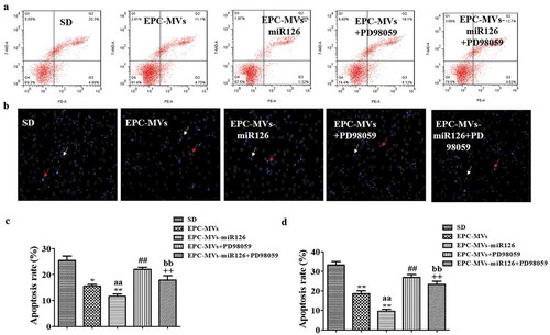
Figure 6. The effects of EPC-MVs and EPC-MVs-miR126 on Erk1/2-Bcl2 pathway. (a) EPC-MVs group and EPC-MVs-miR126 group increase the expression of Bcl-2 gene in MC3T3-E1 cell. (b) Expression of p-Erk1/2/Erk1/2. (c) Expression of Bcl-2. *p< 0.05 vs. control; ##p< 0.01 vs. EPC-MVs; +p< 0.05, ++p< 0.01 vs. EPC-MVs-miR126; aap<0.01 vs. EPC-MVs; bbp<0.01 vs. EPC-MVs+PD98059.
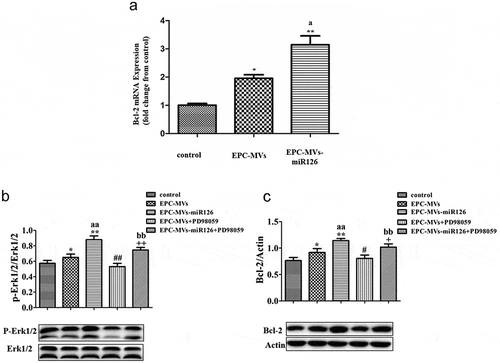
Figure 7. The effects of EPC-MVs and EPC-MVs-miR126 on osteogenic differentiation in MC3T3-E1 cell. (a) The mineralized nodules were stained by Alizarin Red S. (b) The mineralization was quantified with 10% CPC. (c) The mRNA expression of osteogenic genes (Runx2, ALP and OPN) at day 14.
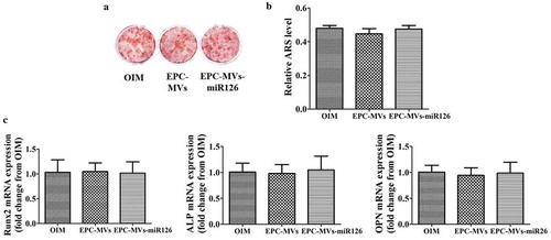
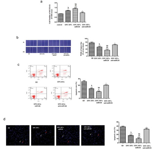
Figure 8. Effects of knock-down of miR126 on MC3T3-E1 cells. (a) The proliferation of MC3T3-E1 cell in different groups. (b) Representative image of cell migration and summarized data on cell migration. (c) Cell apoptosis was measured by flow cytometry. (d) Summarized data on apoptosis rate (%) assessed Hoechst 33258 staining. *p< 0.05, **p< 0.01 vs. SD; ##p< 0.01 vs. EPC-MVs; ++p< 0.01 vs. EPC-MVs-miR126; aap<0.01 vs. EPC-MVs; bbp<0.01 vs. EPC-MVs-anti-miR126.