
Figures & data
Figure 1. DNA sequencing of the PRNP gene.
Compared with the standard sequence, the substitution of cytosine with adenine at codon 196 of the PRNP gene results in the substitution of alanine for glutamate (E196A). The PRNP polymorphism of codon 129 was 129M/M homozygous.
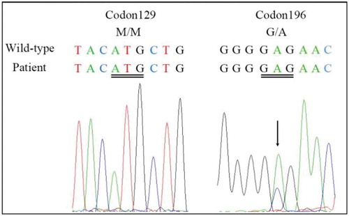
Figure 2. Results of DWI scans.
a and b show an increase in signal intensity changes at the surface of the temporal lobe and bilateral basal ganglia regions (especially the caudate nucleus). c and d show ribbon-like signals in the right parietal lobe.
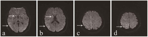
Figure 3. Results of the second EEG.
The second EEG was performed at 2 months after onset of the movement disorder, and a week after the first EEG. The results of the two EEG examinations were similar. a demonstrates abnormal waves in the background. b displays sharp waves, sharp slow waves, and slow waves that are discharged synchronously or asynchronously at the bilateral occipital and frontal region.
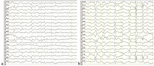
Table 1. Clinical characteristics of the four patients with E196A gCJD.