Figures & data
Figure 1. MRI, single photon emission computed tomography (SPECT) and electroencephalography of the patient. (a): Diffusion-weighted MRI (DW-MRI), and (b): SPECT images obtained 55 months after onset; (c): Electroencephalography obtained 50 months after onset; (d): DW-MRI, and E: SPECT images obtained 55 months after onset; (f): T1-weighted MRI obtained 68 months after onset. The eZIS analysis of SPECT images revealed decreased regional cerebral blood flow (rCBF). A higher Z-score indicated a lower rCBF. A Z-score of 2 to 6 is indicated by the green or black-to-red (lower rCBF) colour gradient. Panel A shows the bilateral hyperintense lesions in the frontal, temporal, and parietal cortices, and the basal ganglia. Panel B depicts the decreased rCBF in the frontal and parietal lobes. Panel C depicts the periodic sharp wave complexes. Panel D depicts bilateral hyperintensity in all cortices, and panel E shows preserved rCBF in the brainstem. Panel F depicts progressive cerebral atrophy, but no brainstem atrophy
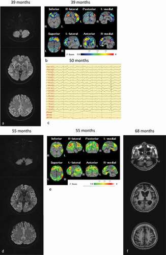
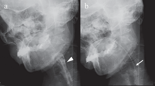
Figure 2. Videofluoroscopic examination of swallowing (VF) of the patient. VF of the patient in frontal view acquired 68 months after the onset of symptoms. (a) Once the swallowing reflex was triggered, the bolus passed through the pharynx into the upper oesophagus (arrowhead). (b) Pharyngeal residue or aspiration was not observed after swallowing (arrow)