Figures & data
Figure 1. Placental homogenates taken from four unique sections of placenta with eight replicates each. There was significant positivity (p = 0.0153) in one section compared to control placenta (star). Positive control CWD+ Muntjac placenta and negative control Muntjac placenta were run concurrently with the assay. sCJD, sporadic Creutzfeldt–Jakob Disease; Norm, Normal control; Plac, placenta; CWD, chronic wasting disease; Neg, negative control; MJ, Muntjac
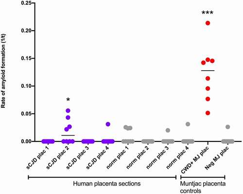
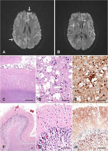
Figure 2. Neuroimaging and brain pathology. (A&B) Diffusion weighted imaging demonstrating cortical ribboning (solid arrow) and restricted diffusion in caudate and putamen (dotted arrow). Occipital neocortex (c-e) and cerebellum vermis (f-h) showing spongiosis with large coalescent vacuoles in the neocortex (c and d), and coarse perivacuolar PrP deposits (e), consistent with the MM2 phenotype of sporadic CJD. The cerebellum shows atrophy of the molecular layer, loss of internal granular layer cells (f and g), and synaptic PrP deposits in the molecular layer (h). C, D, F and G are haematoxylin and eosin stains; E and H are 12F10 prion protein immunolabelled (Cayman Chemical). The size bars are 500 µm (c) and 50 µm (d-h)