Figures & data
Figure 1. Neuropathological finding showing changes corresponding to Creutzfeldt-Jakob disease: (a) Diffuse synaptic positivity and enhanced perivacuolar ‘patchy’ positivity and ‘plaque-like’ positive structures in cerebral cortex, stained with monoclonal antibody (12F10) against prion protein, original magnification 200. (b-c) Diffuse synaptic positivity and enhanced perivacuolar ‘patchy’ positivity and ‘plaque-like’ positive structures in striatum, stained with monoclonal antibody (6H4) against prion protein, original magnification 200x and H&E staining, original magnification 100x
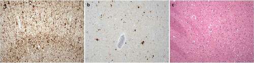
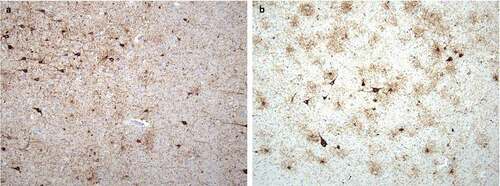
Figure 2. Neuropathological finding corresponding to concomitant tauopathies: (a) Deposits of hyperphosphorylated tau protein in the amygdala corresponding to primary age-related tauopathy (PART); staining with monoclonal antibody against hyperphosphorylated tau (AT8), original magnification 100x. (b) Deposits of hyperphosphorylated tau protein in the amygdala corresponding to age-related tau astrogliopathy (ARTAG); staining with monoclonal antibody against hyperphosphorylated tau (AT8), original magnification 100x