
Figures & data
Figure 1. Brain computed tomography image of an 84-year-old woman shows mild cortical atrophy in the hippocampus and the absence of a cerebrovascular lesion.
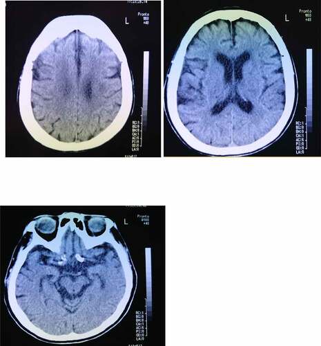
Figure 2. a. Magnetic resonance imaging (MRI) findings obtained using fluid-attenuated inversion-recovery (FLAIR) (upper) and diffusion-weighted images (DWI) (lower) in an 89-year-old woman. DWI shows cortical hyperintensities in some areas of both hemispheres; the white arrows indicate areas of cortical hyperintensity in the left hemisphere.
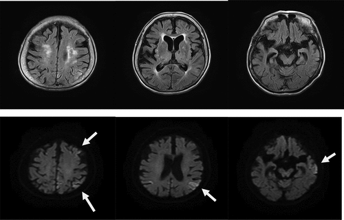
Figure 2. b. Magnetic resonance imaging (MRI) findings obtained using fluid-attenuated inversion-recovery (FLAIR) (upper) and diffusion-weighted images (DWI) (lower) in an 89-year-old woman at 4 months after the initial examination. DWI shows the expansion of the cortical hyperintensity areas in both hemispheres, relative to the previous findings.
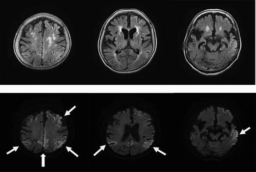
Figure 3. Clinical course of the present case with V180I CJD. In the present case, person DMS was first observed at the age of 84 years. The cognitive impairment progressed to motivation loss and sensory aphasia at the age of 89 years. The patient died because of aspiration pneumonia at the age of 90 years.
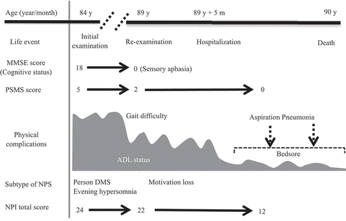
Table 1. Initial NPSs in cases with V180I genetic mutation CJD