
Figures & data
Figure 1. Pedigree of this case. gCJD, genetic Creutzfeldt–Jakob disease; AO, age of onset (year); DD, duration from onset to death (year). Circles indicate females; squares indicate males; yellow symbols indicate affected individuals; diagonal bars indicate deceased members; black arrow indicates the pro-band.
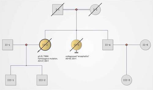
Figure 2. Timeline of the clinical manifestations and the results of examinations of this case. MRI, magnetic resonance imaging; DWI, diffusion-weighted imaging; FDG-PET, 18Fluorodeoxyglucose-positron emission tomography; EEG, electroencephalography.
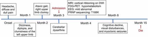
Figure 3. (a) Axial serial diffusion-weighted MRI showing restricted diffusion involving the right striatum, cerebellar cortex and cortical ribboning of the temporal and insular cortices; (b) 18Fluorodeoxyglucose-positron emission tomography showing severe glucose hypometabolism in the bilateral cerebellar cortex and mild glucose hypometabolism in the right frontoparietal cortex; (c) PRNP sequencing showing a homozygous substitution: c.563 C > A (p.T188K).
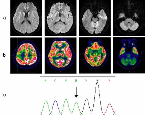
Table 1. Clinical and investigational features of patients with homozygous mutations in the PRNP.