Figures & data
Figure 1. Family tree of the patient. The proband in this family was the present patient (1b). Her father (2a) used to have similar symptoms and dead at 60 years old. Her daughter (1c) have no similar symptom up to now.
Table 1. Clinical data of the patient’s family.
Figure 2. Brain MRI images of the patient (acquired 11-04-2018). (a) Isointensity on T1-weighted fluid-attenuated inversion recovery. (b) Right side of the parietal cortex, T2-weighted fluid-attenuated inversion recovery, slightly high signal. (c, d) Right side of the parietal cortex, diffusion-weighted imaging, high signal.
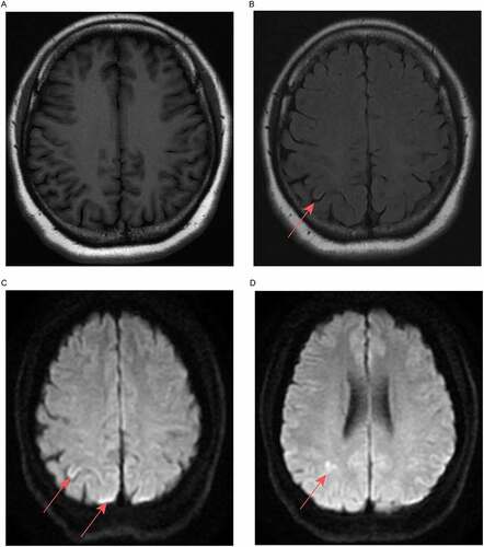
Figure 3. EEG results of the patient. The results showed diffuse slow waves with sharp waves and three-phase waves.
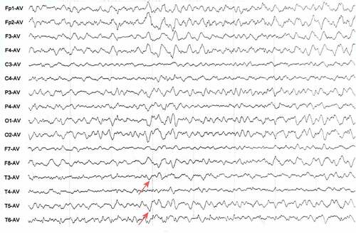
Figure 4. Gene mapping of the patient (performed on 17-05-2018). A missense mutation (C to T) was identified at nt 305 in one allele of PRNP, which led to a change from proline (Pro) to leucine (Leu) at codon 102.
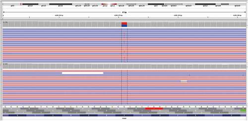
Figure 5. EEG results of the patient after 2 months of TCM treatments. EEG showed diffuse slow waves without sharp waves.