
Figures & data
Figure 1. Human CBs contain cells that express PrPC. CBs are identified with haematoxylin and eosin (Panel a) and toluidine blue (Panel b) and consist of clusters of cells separated by bands of connective tissue. Note the large number of blood vessels that supply the clusters of cells; lumens of some blood vessels are indicated by arrows. Cells in the CBs that express PrPC are not Type I or Type II cells, but instead are relatively large oval or oblong cells scattered throughout the CB and located primarily in the connective tissue that surrounds the CB lobules (Panel c); lumen of some blood vessels are indicated by arrows. A single PrPC-expressing cell (inside the box in Panel c) is enlarged in Panel d and is seen to have the morphology and dense granular appearance that is characteristic of mast cells; lumens of some blood vessels are indicated by arrows. Scale bars: Panel a,b, c = 100 µm; Panel d = 10 µm.
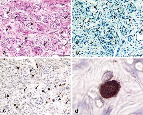
Figure 2. Mast cells in connective tissue septa of CBs can be identified with alcian blue/nuclear fast red stain (Panels a, b) or an antibody against mast cell tryptase (Panels c, d). Note the distribution, morphology, and size of cells stained with alcian blue is similar to the distribution, morphology, and size of the cells stained with the rabbit polyclonal antibody generated against mast cell tryptase. The cell located in the box of panel a and enlarged in panel B is similar to the cell located in the box of panel c that is enlarged in panel d. The mast cells indicated in this figure possess the same distribution, morphology and size as the PrPC-expressing cells shown in panels c and d of figure 1. Scale bars: Panels a, c = 50 µm; Panels b, d = 10 µm.
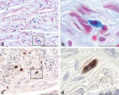
Figure 3. The cells expressing PrPC in the human carotid body are mast cells. Tissue sections of human CB immunohistochemically stained for PrPC expression and counterstained with either TB/ammonium sulphate (Panels a, b) or alcian blue/nuclear fast red (Panels c, d) demonstrate that mast cells express PrPC. PrPC expression is indicated by the brown reaction product, while the mast cell phenotype is indicated by purple (panel b) or blue (panel d) staining of the cell cytoplasm. The colocalization of the two markers in the same cell indicate that mast cells express PrPC. Scale bars: Panels a, c = 100 µm; Panels b, d = 10 µm.
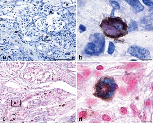
Table 1. Donor body information and tissue collection.
Figure 4. PrPC-expressing mast cells in the human carotid body are located within microns of blood vessels (indicated by asterisks) and neural elements (stained brown). Mast cells that express PrPC can be seen near type I CB cells that contain presynaptic vesicles stained with an antibody against synaptophysin (panel a) and axons stained with an antibody against neurofilament-l (panel b). Type I cells in the CB are innervated by peripheral nerve terminals of the carotid sinus nerve; most of the axons located in the CB are components of the carotid sinus nerve. The carotid sinus nerve terminates in the nucleus of the solitary tract in the brainstem, a known clinical target area for prion neuroinvasion. The proximity of the PrPC-expressing cells, permeable blood vessels and neural elements is consistent with the potential for prion neuroinvasion when there is a prionemia. Scale bars: Panels a, b = 20 µm.
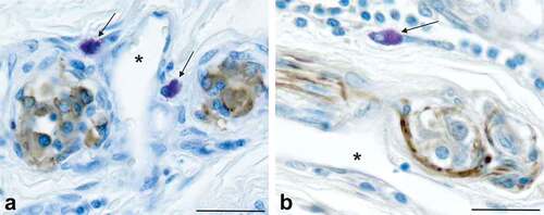
Figure 5. Schematic representation of the potential routes of neuroinvasion following carotid body exposure to prions in blood. The circles represent the location of neuronal cell bodies, the lines represent axons, and the ‘v’ line splits represent axon terminals. The carotid body is located near the bifurcation of the common carotid artery. It is a highly perfused sensory structure that is densely innervated by sensory fibres of the carotid sinus nerve (a branch of the glossopharyngeal nerve) and postganglionic fibres of sympathetic neurons whose cell bodies are located in the superior cervical ganglion. These neural pathways are synaptically linked to areas of the CNS known to be affected early in prion neuroinvasion: the nucleus of the solitary tract (NTS), the dorsal motor nucleus of the vagus (DMNV) and the intermediolateral cell column of the thoracic spinal cord (IML). These structures have been identified as early sites of prion neuroinvasion following oral exposure; note the similarity of this figure to Figure 5 in [Citation33] and Figure 3 in [Citation50]. This figure was created with BioRender.Com.
![Figure 5. Schematic representation of the potential routes of neuroinvasion following carotid body exposure to prions in blood. The circles represent the location of neuronal cell bodies, the lines represent axons, and the ‘v’ line splits represent axon terminals. The carotid body is located near the bifurcation of the common carotid artery. It is a highly perfused sensory structure that is densely innervated by sensory fibres of the carotid sinus nerve (a branch of the glossopharyngeal nerve) and postganglionic fibres of sympathetic neurons whose cell bodies are located in the superior cervical ganglion. These neural pathways are synaptically linked to areas of the CNS known to be affected early in prion neuroinvasion: the nucleus of the solitary tract (NTS), the dorsal motor nucleus of the vagus (DMNV) and the intermediolateral cell column of the thoracic spinal cord (IML). These structures have been identified as early sites of prion neuroinvasion following oral exposure; note the similarity of this figure to Figure 5 in [Citation33] and Figure 3 in [Citation50]. This figure was created with BioRender.Com.](/cms/asset/d1a2fd08-eb8a-40ff-947e-7b52bb622b87/kprn_a_2193128_f0005_oc.jpg)
Table 2. Antibodies and isotype controls.