1,652
Views
0
CrossRef citations to date
0
Altmetric
Research paper

Cerebral cortex swelling in V180I genetic Creutzfeldt–Jakob disease: comparative imaging study between sporadic and V180I genetic Creutzfeldt–Jakob disease in the early stage

, ORCID Icon, , , , , , , & show all
Pages 105-110 | Received 21 Jan 2023, Accepted 14 Mar 2023, Published online: 04 Apr 2023

Figures & data

Table 1. Comparison of the clinical features between V180I and sporadic Creutzfeldt-Jakob disease (sCJD).

Figure 1. Magnetic resonance imaging (MRI) of an 80-year-old male with V180I (a – d) and an 84-year-old male with sporadic Creutzfeldt – Jakob disease (sCJD) (e, f). Brain MRI revealed increased signal intensity in the cerebral cortices on diffusion-weighted imaging in patients with both V180I CJD (a, c) and sCJD (e). Increased signal intensity in the cerebral cortices was also observed on T2-weighted images, with swelling in one patient with V180I CJD (b, d) and no swelling in one patient with sCJD (f).

Figure 1. Magnetic resonance imaging (MRI) of an 80-year-old male with V180I (a – d) and an 84-year-old male with sporadic Creutzfeldt – Jakob disease (sCJD) (e, f). Brain MRI revealed increased signal intensity in the cerebral cortices on diffusion-weighted imaging in patients with both V180I CJD (a, c) and sCJD (e). Increased signal intensity in the cerebral cortices was also observed on T2-weighted images, with swelling in one patient with V180I CJD (b, d) and no swelling in one patient with sCJD (f).

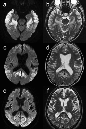
Table 2. Frequency of grey matter hyperintensities at the gyral and nuclear levels in patients with V180I and sporadic Creutzfeldt-Jakob disease (sCJD) (regions with p < 0.1).

Supplemental material

Supplemental Material

Download MS Excel (17 KB)