
Figures & data
Figure 1. Neuroimages, electroencephalographic findings and paraneoplastic panel of the subject. (A) Serum paraneoplastic panel showed a positive band in anti-recoverin autoantibody region. (B) Electroencephalography showing nearly continuous generalized quasiperiodic sharp slow or sharp-and slow waves at 1–2 Hz, 50–100 uV, with emphasis in posterior head areas. (C-D) Axial view of brain magnetic resonance imaging diffusion weighted imaging showing hyperintensity over bilateral parieto-temporo-occipital lobes (arrowheads) and bilateral putamen and caudate nuclei (arrow), but no contrast enhancement in T1-weighted image.
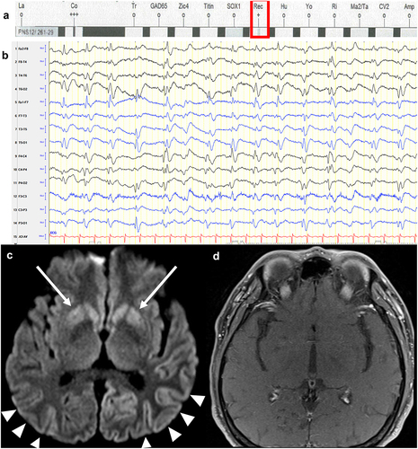
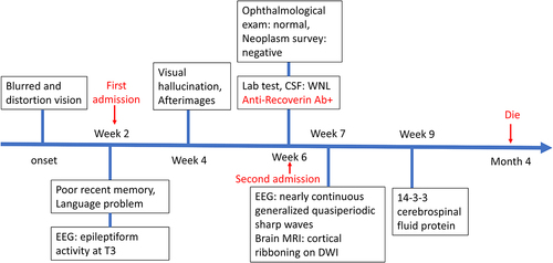
Figure 2. Timeline of the clinical manifestations and the results of examinations of the present case. Ab, antibody; CSF, cerebrospinal fluid DWI, diffusion-weighted imaging; EEG, electroencephalography; MRI, magnetic resonance imaging; WNL, within normal limit.
Data availability statement
The complete data are available from the corresponding author on reasonable request.