Figures & data
Figure 1. Effects of BJhTERT fibroblasts on proliferation of normal and cancer prostate epithelial cells in vitro. (A) Morphology of cocultures at day 1 and day 2 (light microscopy), magnification ×200. (B) Proliferation rates of normal (PNT2) and cancer epithelial prostate cells before and after coculture with fibroblasts (CyQUANT NF Cell proliferation assay).
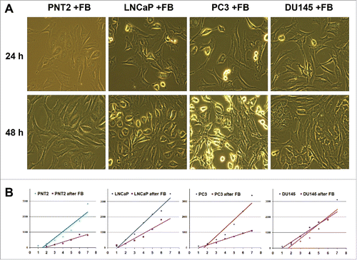
Figure 2. Fibroblast coculture effect on normal and cancer prostate epithelial cells. Changes in expression of proteoglycans (A) and ECM components (B) in normal and cancer prostate cells upon coculture with fibroblasts. Intensity of the amplified DNA fragments normalized to that of GAPDH.
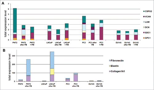
Figure 3. Effects of normal and cancer prostate epithelial cells to TERT-immortilized fibroblasts. (A) Proteoglycan expression. (B) ECM components expression. (C) Changes in fibroblast proliferation rate upon coculture with prostate epithelialcells.
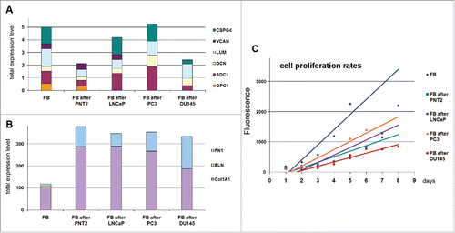
Figure 4. Immunocytochemical analysis of proteoglycans expression in fibroblasts (FB) and normal (PNT2) and cancer prostate cells (LNCaP, PC3, DU145) before and after coculture with fibroblasts. Decorin (green), lumican (red), syndecan-1 (green), glypican-1 (red). Nuclear counterstain - DAPI (blue).
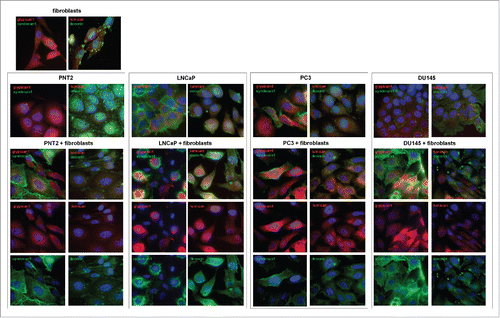
Figure 5. RT2 Profiler™ PCR Array Human Cell Junction Pathway Finder analysis of normal and cancer prostate epithelial cells before and after coculture with BJhTERT fibroblast. (A) The relative expression levels for each gene in PNT2 cells (Group 1) or PC3 cells (Group 3) after co-culture with fibroblasts are plotted against the same gene from the control PNT2 cells or PC3 cells (Control Group or Group 2, respectively). The middle line shows the similar expression in both groups with 2-fold change boundaries. Genes up-regulated >2-fold lie above the middle line and the down-regulated genes lie below the line. (B) Gene expression levels for PNT2 cells (Control group), BJhTERT-exposed PNT2 cells (Group 1), PC3 cells (Group 2) and BJhTERT-exposed PC3 cells (Group 3) (RT2 Profiler™ PCR Array Data Analysis Program version 3.5).
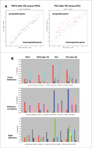
Table 1. Changes in the expression of genes relevant to the Cell Junction pathway. Listed up- and down-regulated genes in the Test Sample (after coculture) divided by the normalized gene expression in the Control Sample (before coculture). Two-fold change is taken as significant.
Figure 6. Expression profiling of cell junction-related genes of BJhTERT fibroblasts before and after coculture with normal and cancer prostate epithelial cells. (A) RT2 Profiler™ PCR Array Human Cell Junction Pathway Finder analysis. The relative expression levels for each gene in fibroblasts after co-culture with PNT2 cells (Group 1) or PC3 cells (Group 2) are plotted against the same gene from the control fibroblasts (Control Group). The middle line shows the similar expression in both groups with 2-fold change boundaries. Genes upregulated >2-fold lie above the middle line and the downregulated genes lie below the line. (B) Gene expression levels for fibroblasts, PNT2- and PC3-confronted fibroblasts for junctions pathways with highest expression levels. (C) Immunostaining for a-smooth muscle actin (SMA) (green). Nuclear counterstain - DAPI (blue).
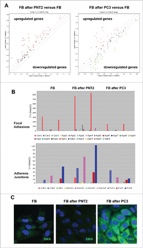
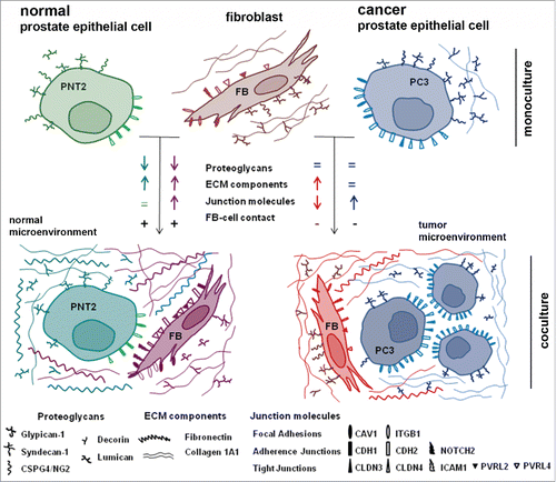
Figure 7. Schematic representation of a running hypothesis for fibroblast – prostate epithelial cells interactions.