Figures & data
Figure 1. A diagram representing the structure of capsaicinoids and capsaicin-analogs. (A) General structure of capsaicinoids. R = alkyl group. (B) Molecular structure of arvanil, olvanil and capsaicin.
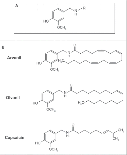
Figure 2. Diagrammatic illustration of the spherical invasion assay. (A) The first layer is comprised of DMS 114 cells mixed in a 1:1 suspension with phenol-red containing Matrigel (light pink area). After 18 hours, DMS 114 cells grow and extend up to the boundary of this first layer. (B) After 24 hours, a second layer of 1:1 solution phenol-red free Matrigel, in phenol-red free RPMI (gray area) is added on top of the first Matrigel spot. The cells are incubated for 24 hours at 37°C. (C) After 24 hours, DMS 114 cells invade into the secondary Matrigel layer. The chamber slides are observed by phase contrast microscopy. (D) A representative photograph of untreated DMS 114 cells is shown. The black arrow indicates the vessels in second layer. The dotted line ab is taken as the interface between the two layers. The distance to which the cells have traveled (into the secondary Matrigel layer) is measured at seven sites (for each photograph) in a randomized double blind fashion by three independent observers, using NIH Image J Version 1.47. This process is repeated for three separate photographic fields per sample.
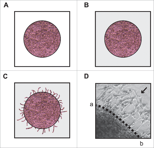
Figure 3. Capsaicin suppresses invasion of DMS 114 human SCLC cells in a concentration-dependent and time-dependent manner. (A) Boyden chamber assays indicate that capsaicin inhibited the invasion of DMS 114 human SCLC cells in a concentration-dependent manner over 24 hours. (B) MTT assays show that capsaicin decreases the viability of DMS 114 cells from concentrations ranging from 30 μM-100 μM over 24 hours. (C) Time kinetics of the anti-invasive activity of 20 μM capsaicin from 24–48 hours. Values indicated by * are statistically significant relative to controls. The absorbance of control cells was assumed to be 1, and arvanil-, olvanil-, capsaicin-induced decreases in invasion were calculated as fold change from control. (D) MTT assays show that 20 μM capsaicin does not impact the viability of DMS 114 cells at 24 hours or 36 hours. However, the treatment of 20 μM capsaicin decreases cell viability in DMS 114 cells over 48 hours. The absorbance of control cells was assumed to be 1, and arvanil-, olvanil-, capsaicin-induced decreases in cell viability were calculated as fold change from control. Values indicated by * are statistically significant relative to controls. The figure represents the average of two independent experiments, and the data have been represented as mean ± SEM.
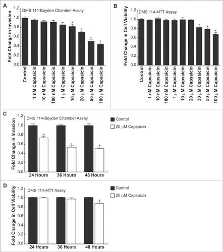
Figure 4. (see previous page) Arvanil and olvanil decrease the invasion of DMS 114 human SCLC cells (A) Boyden chamber assays indicate that arvanil inhibited the invasion of DMS 114 human SCLC cells in a concentration-dependent manner over 24 hours. Arvanil displayed significant anti-invasive activity at 10 nM-100 μM (P < 0 .05) (B) Olvanil suppressed the invasion of DMS 114 human SCLC cells in a concentration-dependent manner. Olvanil decreased the invasion of DMS 114 cells between 10 nM-100 μM. The absorbance of control cells was assumed to be 1, and arvanil-, olvanil-, capsaicin-induced decreases in invasion were calculated as fold change from control. (C) MTT assays reveal that arvanil and olvanil decreased the viability of DMS 114 cells at concentrations ranging from 10 μM-100 μM. Values indicated by * are statistically significant relative to controls. Based on the combined results of the Boyden chamber assay and MTT assay, the concentrations of 1 μM arvanil, 1 μM olvanil and 20 μM capsaicin were chosen for subsequent experiments. The absorbance of control cells was assumed to be 1, and arvanil-, olvanil-, capsaicin-induced decreases in cell viability were calculated as fold change from control. (D) The Boyden chamber assays were repeated in a second human SCLC cell line DMS 53 and similar results were obtained. (E) MTT experiments indicate that 1 μM arvanil, 1 μM olvanil and 20 μM capsaicin do not cause any change in the viability of DMS 53 cells over 24 hours. The absorbance of control cells was assumed to be 1, and arvanil-, olvanil-, capsaicin-induced decreases in invasion were calculated as fold change from control. Values indicated by same letter are not statistically significant. The figure represents the average of two independent experiments, and the data have been represented as mean ± SEM.
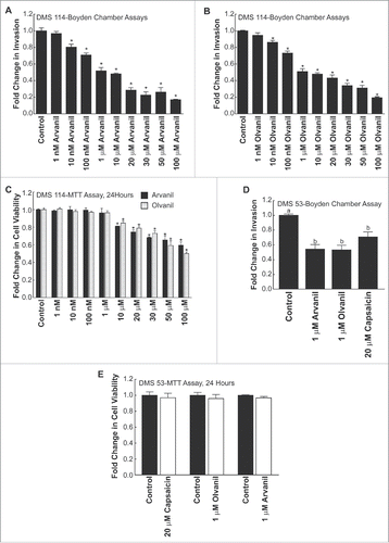
Figure 5. (see previous page) Arvanil, olvanil and capsaicin suppress invasion of DMS 114 cells in spherical invasion assay. (A) Spherical invasion assays indicate that untreated DMS 114 showed robust invasion of cells (indicated by black arrows) into the secondary Matrigel layer across the interface ab (Left column, top, middle and bottom pictures). The treatment of DMS 114 cells with 1 μM arvanil (Right column, top picture) or 1 μM olvanil (Right column, middle picture) or 20 μM capsaicin (Right column, bottom picture) caused a substantial inhibition of invading DMS 114 cells (indicated by black arrows) across the line ab in the secondary Matrigel layer. (B) Quantification of the anti-invasive activity of 1 μM arvanil, 1 μM olvanil and 20 μM capsaicin in DMS 114 cells, as measured by the spherical invasion assay. (C) The experiment was repeated in DMS 53 human SCLC cells, and similar results were obtained. The distance traveled by the control cells was assumed to be 1, and arvanil-, olvanil-, capsaicin-induced decreases in invasion were calculated as fold change from control. Values indicated by the same letter are not statistically significant. The figure represents the average of two independent experiments and the data have been represented as mean ± SEM. (D) MTT assays demonstrate that 1 μM arvanil or 1 μM olvanil or 20 μM capsaicin do not decrease the viability of DMS 114 cells (black bars) or DMS 53 cells (white bars) over 36 hours. The absorbance of control cells was assumed to be 1, and arvanil-, olvanil-, capsaicin-induced decreases in cell viability were calculated as fold change from control. The figure represents the average of two independent experiments, and the data have been represented as mean ± SEM.
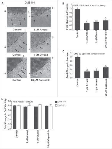
Figure 6. The anti-invasive activity of arvanil, olvanil and capsaicin were independent of TRPV pathway, CB1 pathway and required AMPK activation in DMS 114 cells. (A) Boyden chamber assays reveal that the anti-invasive activity of arvanil, olvanil and capsaicin were unaffected by the generalized TRPV antagonist ruthenium red in DMS 114 cells. (B) The invasion-inhibitory activity of arvanil, olvanil and capsaicin is not influenced by the CB1 antagonist AM281 in DMS 114 cells, as measured by Boyden chamber assays. (C) The AMPK inhibitor dorsomorphine dichloride reverses the anti-invasive activity of arvanil, olvanil and capsaicin in DMS 114 cells, as analyzed by Boyden chamber invasion assays. The absorbance of control cells was assumed to be 1, and arvanil-, olvanil-, capsaicin-induced decreases in invasion were calculated as fold change from control. Values indicated by the same letter are not statistically significant. The figure represents the average of two independent experiments and the data have been represented as mean ± SEM.
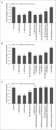
Figure 7. Spherical invasion assays reveal that the anti-invasive activity of arvanil, olvanil and capsaicin did not involve the TRPV or CB1 pathway but were mediated by the AMPK pathway, in DMS 114 cells (A) Spherical invasion assays demonstrate that the anti-invasive activity of arvanil, olvanil or capsaicin were unaffected by the generalized TRPV antagonist ruthenium red in DMS 114 cells. (B) Spherical invasion assays reveal that invasion-inhibitory activity of arvanil, olvanil and capsaicin was not abrogated by the CB1 antagonist AM281 in DMS 114 cells. (C) The AMPK inhibitor dorsomorphine dichloride reverses the anti-invasive activity of arvanil, olvanil and capsaicin in DMS 114 cells, as measured by spherical invasion assays. The distance traveled by the control cells was assumed to be 1, and arvanil-, olvanil-, capsaicin-induced decreases in invasion were calculated as fold change from control. Values indicated by the same letter are not statistically significant. The figure represents the average of two independent experiments and the data have been represented as mean ± SEM.
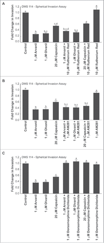
Figure 8. Human SCLC cells express AMPK-α1 but not AMPK-α2. Arvanil, olvanil and capsaicin block activation of AMPK-α1 in human SCLC cells. (A) Immunoblotting experiments show that AMPK-α1 is robustly expressed in DMS 114 and DMS 53 cells. T47D human breast cancer cells were used as the positive controls for the experiment. GAPDH was used as the loading control for the protein gel blotting experiments, and the results were quantitated by densitometric analysis. (B) Western blotting experiments demonstrate that AMPK-α2 is not expressed by DMS 53 and DMS 114 human SCLC cells. T47D human breast cancer cells were used as the positive controls for the experiment. GAPDH was used as the loading control for the western blotting experiments, and the results were quantitated by densitometric analysis. (C) The treatment of 1 μM arvanil caused robust phosphorylation (and activation) of AMPK-α1 at threonine residue 172, in DMS 114 and DMS 53 cells over 24 hours (top panel). Total AMPK levels remained constant (middle panel). GAPDH was used as the loading control (bottom panel) for the protein gel blotting experiments, and the results were quantitated by densitometric analysis. The experiments were repeated using 1 μM olvanil (D) and 20 μM capsaicin (E) and similar results were obtained.
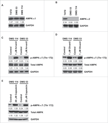
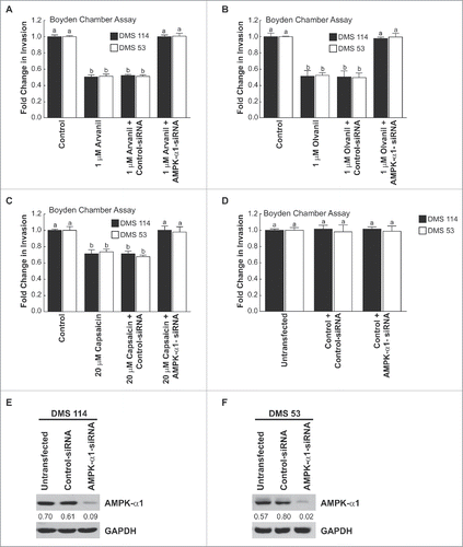
Figure 9. The AMPK-pathway mediated the anti-invasive activity of arvanil, olvanil and capsaicin in human SCLC cells. (A) Boyden chamber assays indicated that the transfection of AMPK-α1-siRNA significantly abrogated the anti-invasive activity of arvanil in DMS 114 cells (black bars). The experiment was repeated in human DMS 53 cells and comparable results were obtained (white bars). The transfection of a non-targeting control-siRNA did not have any effect on the anti-invasive activity of arvanil in DMS 114 and DMS 53 cells. (B) Depletion of AMPK-α1 levels by siRNA techniques ablated the anti-invasive activity of olvanil in DMS 114 (black bars) and DMS 53 cells (white bars). The anti-invasive activity of olvanil was not influenced by transfection of a control non-targeting siRNA in DMS 114 and DMS 53 cells. (C) AMPK-α1-siRNA reversed the anti-invasive activity of capsaicin in DMS 114 (black bars) and DMS 53 cells (white bars), whereas the control-siRNA have not effect on the anti-invasive activity of capsaicin in these two cell lines. The absorbance of control cells was assumed to be 1, and arvanil-, olvanil-, capsaicin-induced decreases in invasion were calculated as fold change from control. (D) The transfection of AMPK-α1-siRNA did not have any effect on the invasion of untreated DMS 114 (black bars) or DMS 53 cells (white bars). (E) Western blotting analysis showed that AMPK-α1 expression in DMS 114 cells and DMS 53 cells (F) was suppressed upon siRNA transfection. GAPDH was used as the loading control for the western blotting experiments, and the results were quantitated by densitometric analysis.