Figures & data
Figure 1. TGF-β triggers the in vitro motility of osteosarcoma cells. (A) MG-63 and HOS cells were treated with or without TGF-β (20 ng/ml) and then scraped by a pipette tip to generate wounds for 48 h, representative images of wounds were observed; (B) The statistic results of wound healing assays; (C) MG-63 and HOS cells treated with or without TGF-β (20 ng/ml) were allowed to invade transwell chambers for 48 h, then the invaded cells were fixed, stained, and counted. Data are presented as means ± SD of 3 independent experiments. **p < 0.01 compared with control.
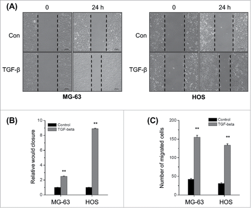
Figure 2. TGF-β triggers the EMT of osteosarcoma cells. (A) MG-63 and HOS cells were treated with or without TGF-β (20 ng/ml) for 48 h, then the protein levels of E-cad, ZO-1, Vim, and FN were analyzed by Western blot analysis; (B) MG-63 and HOS cells were treated with or without TGF-β (20 ng/ml) for 24 h, then the mRNA levels of E-cad, ZO-1, Vim, and FN were analyzed by qRT-PCR. Data are presented as means ± SD of 3 independent experiments. **p < 0.01 compared with control.
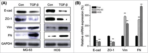
Figure 3. Up regulation of Snail mediates TFG-β induced EMT of osteosarcoma cells. MG-63 and HOS cells were treated with TFG-β (20 ng/ml) for 24 h, the protein (A) and mRNA (B) levels of Snail, Slug, Twist, and ZEB were analyzed by Western blot analysis or qRT-PCR, respectively; (C) MG-63 cells were transfected with Snail specific si-RNA (si-Snail) or negative control si-RNA (si-NC) for 24 h, and then the mRNA of Snail were analyzed by qRT-PCR; (D) MG-63 cells transfected with si-Snail or si-NC were stimulated with or without TFG-β (20 ng/ml) for 48 h, the protein levels of Snail, E-Cad, and Vim were analyzed by Western blot analysis. Data were presented as means ± SD of 3 independent experiments. **p < 0.01 compared with control.
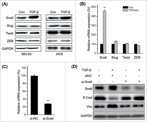
Figure 4. TGF-β increases the expression and nuclear localization of ERRα in osteosarcoma cells. (A) MG-63 and HOS cells were treated with or without TGF-β (20 ng/ml) for 24 h, and then the express of protein (A) and mRNA (B) of ERRα, ERα, ERβ, or ERRγ were measured by use of Western blot analysis and qRT-PCR, respectively; (C) MG-63 cells were treated with or without TGF-β (20 ng/ml) for 24 h, the cellular location of ERRα (green) were examined by immunofluorescence staining and nuclei were stained with DAPI (blue). The quantification results were shown in the right column. Data were presented as means ± SD of 3 independent experiments. **p < 0.01 compared with control. Scale bar = 50 μm.
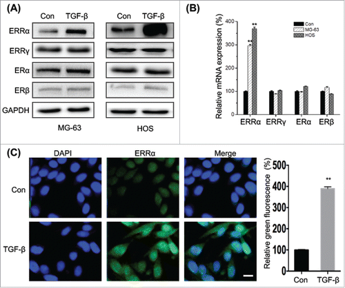
Figure 5. Knock down of ERRα attenuates TFG-β induced EMT of osteosarcoma cells. MG-63 and HOS cells were transfected with si-NC or si-ERRα for 24 h, and then the expression of ERRα was measured by use of Western blot analysis and qRT-PCR; MG-63 (B) and HOS (C) cells were transfected with si-NC or si-ERRα for 24 h and then further treated with TFG-β (20 ng/ml) for 48 h, the protein levels of E-cad and Vim were analyzed by Western blot analysis; (D) MG-63 cells were transfected with si-NC or si-ERRα for 24 h and then further treated with TFG-β (20 ng/ml) for 24 h, the mRNA levels of E-cad and Vim were analyzed by qRT-PCR. Data were presented as means ± SD of 3 independent experiments. **p < 0.01 compared with control.
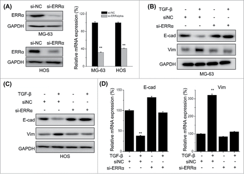
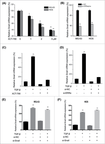
Figure 6. ERRα is involved in TFG-β induced transcription of Snail in osteosarcoma cells. MG-63 and HOS cells were treated with increasing concentrations of XCT-790 (A) or transfected with si-ERRα for 24 h, the expression of Snail mRNA were analyzed by qRT-PCR; (C) MG-63 cells were treated with TFG-β (20 ng/ml), XCT-790 (2 μM) or both of them for 24 h, the expression of Snail mRNA were analyzed by qRT-PCR; (D) MG-63 cells were transfected with si-ERRα or si-NC for 24 h and then treated with TFG-β (20 ng/ml) for 24 h, the expression of Snail mRNA were analyzed by qRT-PCR; MG-63 (E) or HOS (F) cells were transfected with siNC or si-Snail for 24 h and then further treated with TFG-β (20 ng/ml) for another 24 h, the mRNA expression of ERRα were measured by use of qRT-PCR. Data were presented as means ± SD of 3 independent experiments. **p < 0.01 compared with control.