3,225
Views
18
CrossRef citations to date
0
Altmetric
Brief Communication

A standardized nomenclature and atlas of the male terminalia of Drosophila melanogaster

ORCID Icon, , ORCID Icon, ORCID Icon, , , ORCID Icon, ORCID Icon, ORCID Icon, ORCID Icon, ORCID Icon, ORCID Icon, ORCID Icon, ORCID Icon & ORCID Icon show all
Pages 51-64 | Received 17 Jun 2019, Accepted 04 Aug 2019, Published online: 19 Aug 2019

Figures & data

Figure 1. (a) Light microscope preparation of the entire male terminalia of D. melanogaster Canton S. (b) Caudolateral view of the periphallic structures. (c) Ventrolateral view of the phallic structures. Scale bars are 100 μm. Note that the exact size and shape of terminalia structures, such as the epandrial posterior lobe, vary within D. melanogaster [Citation27,Citation28].

Figure 1. (a) Light microscope preparation of the entire male terminalia of D. melanogaster Canton S. (b) Caudolateral view of the periphallic structures. (c) Ventrolateral view of the phallic structures. Scale bars are 100 μm. Note that the exact size and shape of terminalia structures, such as the epandrial posterior lobe, vary within D. melanogaster [Citation27,Citation28].

Table 1. Definition of the terms in the standardized nomenclature.

Table 2. Table of correspondence between terms previously used in publications and term of the standardized nomenclature.
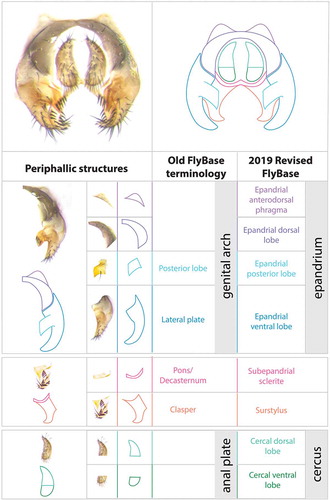
Figure 2. Visual atlas of periphallic structures. Light microscopy images (Canton S strain) and diagrams representing the broad divisions and substructures of epandrium and cercus. The images are oriented dorsal (top) to ventral (bottom). Previous FlyBase terms are on the left and 2019 revised terms are on the right.

Figure 2. Visual atlas of periphallic structures. Light microscopy images (Canton S strain) and diagrams representing the broad divisions and substructures of epandrium and cercus. The images are oriented dorsal (top) to ventral (bottom). Previous FlyBase terms are on the left and 2019 revised terms are on the right.

Figure 3. Visual atlas of phallic structures. Light microscopy images (Canton S strain) and diagrams representing the broad divisions and substructures of phallus and hypandrium. The images are oriented posterior (top) to anterior (bottom). Previous FlyBase terms are on the left and the 2019 revised terms are on the right.

Figure 3. Visual atlas of phallic structures. Light microscopy images (Canton S strain) and diagrams representing the broad divisions and substructures of phallus and hypandrium. The images are oriented posterior (top) to anterior (bottom). Previous FlyBase terms are on the left and the 2019 revised terms are on the right.

Figure 4. Scanning electron micrographs of the phallic structures in (a) ventral and (b) lateral views, from L. Tsacas’ collection at the National Museum of Natural History, Paris (Courtesy of the Museum).

Figure 4. Scanning electron micrographs of the phallic structures in (a) ventral and (b) lateral views, from L. Tsacas’ collection at the National Museum of Natural History, Paris (Courtesy of the Museum).

Figure 5. Musculature of the phallic structures. Same diagram of cuticular parts as in (ventral view). Muscles are indicated in distinct colors and numbered I to VI [Citation18]. These muscles are bilateral. For sake of clarity, muscles are shown either on the left or on the right side of the diagram. See for muscles description.

Figure 5. Musculature of the phallic structures. Same diagram of cuticular parts as in Figure 3 (ventral view). Muscles are indicated in distinct colors and numbered I to VI [Citation18]. These muscles are bilateral. For sake of clarity, muscles are shown either on the left or on the right side of the diagram. See Table 1 for muscles description.

Reprints and Corporate Permissions

Please note: Selecting permissions does not provide access to the full text of the article, please see our help page How do I view content?

To request a reprint or corporate permissions for this article, please click on the relevant link below:

Academic Permissions

Please note: Selecting permissions does not provide access to the full text of the article, please see our help page How do I view content?

Obtain permissions instantly via Rightslink by clicking on the button below:

If you are unable to obtain permissions via Rightslink, please complete and submit this Permissions form. For more information, please visit our Permissions help page.