Figures & data
Figure 1. Posterior crossvein development of the Drosophila pupal wing. (A, A´) During the first apposition of the wing epithelia, around 8 h after pupariation (AP), dpp (green) is expressed in the longitudinal veins 2–5 (LV2-5). (B, B´) In the course of the inflation stage (10 h – 20 h AP), Dpp diffuses laterally to maintain a long-range BMP signal (magenta arrows). (C, C´) During the second apposition stage (20 h AP or later), posterior crossvein (PCV) progenitor cells are first detectable. At this stage, Dpp maintains a short-range BMP signal in the LVs (blue arrows). A long-range BMP signal is needed for PCV formation. (D) A schematic of long-range BMP signal into PCV. Sog/Cv complex facilitates Dpp:Gbb heterodimer trafficking from LVs into the PCV region. Sog is then cleaved by protease Tlr enabling the release of Dpp:Gbb heterodimers, which subsequently activates the Tkv-Punt receptor in the PCV region. Dorsal view of the pupal wing (A, B, C). Cross-section of the area shown in A, B, C with red dashed line (dorsal at the top and ventral at the bottom) (A´, B´, C´). Zoomed view of the PCV region shown in C with black dashed box (D). Created with BioRender.com.
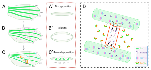
Table 1. Molecular factors involved in the PCV development of the Drosophila pupal wing and their human orthologs
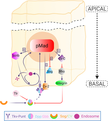
Figure 2. Schematic overview of BMP signalling regulatory system in the PCV cell. The Dpp:Gbb heterodimer is trafficked basally with BMP binding proteins Sog/Cv to bind Tkv-Punt receptor. The release of the ligands requires protease Tlr that is responsible for the cleavage of Sog. The release of the heterodimers leads to the activation of the receptor prompting BMP signalling highlighted by pMad expression. BMP signal regulates various co-factors that form a feedback or feedforward mechanism to further sustain BMP signal in the PCV field. i) Cv-2 is upregulated by BMP signal. Cv-2 is a secreted BMP-binding protein that promotes BMP signalling by facilitating receptor-ligand binding. ii) BMP signal represses sog expression which is needed for continuous PCV formation. iii) BMP signal is needed for up-regulating Scrib expression, which in turn optimizes the BMP signalling by regulating the localization of Tkv and by facilitating Tkv internalization to Rab5 endosomes enabling the receptor signalling. The endosomes can either enter into the receptor recycling or go to degradation. iv) BMP signal induces cv-c expression, which is responsible for inactivating Rho-type small GTPases, leading to the downregulation of β-integrins. Low levels of β-integrins provide optimal extracellular environment to maintain ligand trafficking into the PCV region. Apical portion of the epithelial cell is abbreviated. Created with BioRender.com.