
Figures & data
Figure 1. Phylogeny and distribution of Drosophila elegans and related species. (a) Phylogenetic tree of the melanogaster and related species groups, integrated from robust tree topologies inferred in previous molecular phylogenetic studies [Citation1–5]. (b) Geographic distribution of D. elegans. Texts and filled areas indicate where D. elegans has been collected (light grey for brown morph, dark grey for black morph). Data from DrosWLD-Species (https://bioinfo.museum.hokudai.ac.jp/db/, DATA ID: 76707) [Citation6–12]. A photo shows habitus (dorsal view) of a male of the brown morph type of D. elegans. This is reused with the kind permission of Dr. Nicolas Gompel (Image source: http://gompel.org/wp-content/gallery/drosophilidae/Drosophila-elegans-HK-iso1-genome-male-1x125-dorsal-enhanced.jpg). CC BY-NC-SA 3.0.
![Figure 1. Phylogeny and distribution of Drosophila elegans and related species. (a) Phylogenetic tree of the melanogaster and related species groups, integrated from robust tree topologies inferred in previous molecular phylogenetic studies [Citation1–5]. (b) Geographic distribution of D. elegans. Texts and filled areas indicate where D. elegans has been collected (light grey for brown morph, dark grey for black morph). Data from DrosWLD-Species (https://bioinfo.museum.hokudai.ac.jp/db/, DATA ID: 76707) [Citation6–12]. A photo shows habitus (dorsal view) of a male of the brown morph type of D. elegans. This is reused with the kind permission of Dr. Nicolas Gompel (Image source: http://gompel.org/wp-content/gallery/drosophilidae/Drosophila-elegans-HK-iso1-genome-male-1x125-dorsal-enhanced.jpg). CC BY-NC-SA 3.0.](/cms/asset/9e39f372-6706-4693-8bfd-22e55997c18e/kfly_a_2066953_f0001_oc.jpg)
Figure 2. Morphology of Drosophila elegans brown morph. (a) Male habitus (lateral view). (b) Female habitus (lateral view). (c) Periphallic organs (caudolateral view). (d) Phallic organs (ventral view). (e) Ditto. (f) Female terminalia (lateral view). (g) Ditto (ventral view). (h) Spermatheca (lateral view). Diagnostic characters are indicated with red arrows: (i) aedeagus medially very narrow, apically expanded and truncate with nearly flat margin in ventral view; (ii) posterior elongation of pregonite apically slightly expanded triangularly; (iii) hypogynial valve (oviscapt) with teeth arranged in a single row on ventral margin; and (iv) spermathecal capsule as long as wide, with basal collar. Scales: 1 mm in a and b, 0.1 mm in c–h.
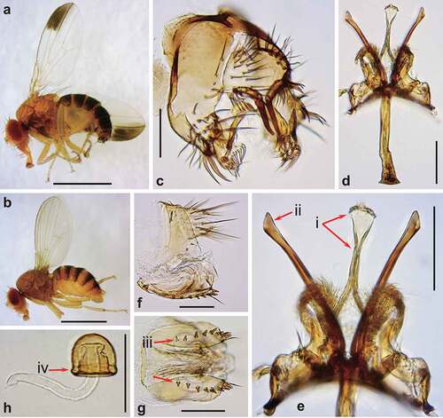
Figure 3. Morphology of Drosophila gunungcola. (a) Male habitus (lateral view). (b) Female habitus (lateral view). (c) Periphallic organs (caudal view). (d) Phallic organs (ventral view). (e) Ditto (lateral view). (f) Female terminalia (lateral view). (g) Ditto (ventral view). (h) Spermatheca (lateral view). Diagnostic characters are indicated with red arrows: (i) aedeagus apically expanded, with convex distal margin in ventral view; (ii) posterior elongation of pregonite apically expanded roundly; (iii) hypogynial valve (oviscapt) with teeth arranged in a few irregular rows on ventral margin; and (iv) spermathecal capsule longer than wide, without basal collar. Scales: 1 mm in a and b, 0.1 mm in c–h.
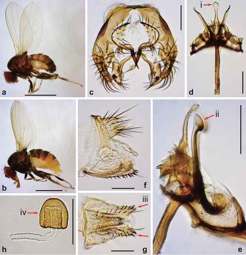
Figure 4. Breeding sites of the Drosophila melanogaster species group, and flower-visiting and courtship behaviours of D. elegans. (a) A phylogenetic tree based on the hypothesis shown in ), with breeding sites of the melanogaster and related species groups [Citation7,Citation10,Citation20–24]. Symbols of fruits and flowers indicate the major breeding sites. Detailed information with references is available in Figure S1. (b) A D. elegans male staying in a flower of the host plant Ipomoea indica (left). The inset shows an enlarged image. A male of D. elegans courting a female with a wing display in a flower of I. indica (right). The inset shows successful copulation of a pair. The photos are captured by Dr. Ryoya Tanaka on Okinawa main Island. (c) D. elegans gathering in a flower of the host plant Alpinia zerumbet. The photos are captured by Dr. Takao Yoshida on Iriomote Island, and shared through Japan Drosophila Database (http://www.drosophila.jp/jdd/index_en.html).
![Figure 4. Breeding sites of the Drosophila melanogaster species group, and flower-visiting and courtship behaviours of D. elegans. (a) A phylogenetic tree based on the hypothesis shown in Figure 1(a), with breeding sites of the melanogaster and related species groups [Citation7,Citation10,Citation20–24]. Symbols of fruits and flowers indicate the major breeding sites. Detailed information with references is available in Figure S1. (b) A D. elegans male staying in a flower of the host plant Ipomoea indica (left). The inset shows an enlarged image. A male of D. elegans courting a female with a wing display in a flower of I. indica (right). The inset shows successful copulation of a pair. The photos are captured by Dr. Ryoya Tanaka on Okinawa main Island. (c) D. elegans gathering in a flower of the host plant Alpinia zerumbet. The photos are captured by Dr. Takao Yoshida on Iriomote Island, and shared through Japan Drosophila Database (http://www.drosophila.jp/jdd/index_en.html).](/cms/asset/88e4791f-e09c-4bb0-aacf-e61abf51bbe0/kfly_a_2066953_f0004_oc.jpg)
Table 1. Tested artificial fly foods varying in ingredient ratios and the breeding performance of Drosophila elegans.
Figure 5. Cuticular hydrocarbons (CHCs) of Drosophila elegans and D. gunungcola. The CHC compositions (%) of black and brown morphs of D. elegans and D. gunungcola. Data from [Citation42,Citation43]. Each point indicates asingle data point from 200 flies.
![Figure 5. Cuticular hydrocarbons (CHCs) of Drosophila elegans and D. gunungcola. The CHC compositions (%) of black and brown morphs of D. elegans and D. gunungcola. Data from [Citation42,Citation43]. Each point indicates asingle data point from 200 flies.](/cms/asset/35587a45-aa53-4cac-835d-084d3270a3f1/kfly_a_2066953_f0005_oc.jpg)
Figure 6. Karyotype and chromosome types of the Drosophila melanogaster and related species groups. A phylogenetic tree with the karyotype and chromosome types [Citation1,Citation13,Citation35,Citation46]. Chromosome types are classified into metacentric (M), submetacentric (sm), subtelocentric (st), telocentric (T), dot (D), and rod-like dot (Dr). In , Muller’s elements A, B, C, D, E, and F correspond to the chromosomes X, 2L, 2R, 3L, 3R, and IV, respectively.
![Figure 6. Karyotype and chromosome types of the Drosophila melanogaster and related species groups. A phylogenetic tree with the karyotype and chromosome types [Citation1,Citation13,Citation35,Citation46]. Chromosome types are classified into metacentric (M), submetacentric (sm), subtelocentric (st), telocentric (T), dot (D), and rod-like dot (Dr). In , Muller’s elements A, B, C, D, E, and F correspond to the chromosomes X, 2L, 2R, 3L, 3R, and IV, respectively.](/cms/asset/d1f25235-2bad-428f-ba38-8bcc8c82fe68/kfly_a_2066953_f0006_b.gif)