Figures & data
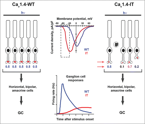
Figure 1. Retinal organization and ganglion cell (GC) signaling in wild-type (Cav1.4-WT) and Cav1.4-IT mice. Compared to WT channels, Cav1.4-IT channels activate at more negative voltages, causing greater Ca2+ influx within the physiological range of PR membrane voltages (dotted rectangle, top graph). In Cav1.4-WT retina (left), temporally precise and uniform PR responses to light rely on normal synapse structure and proper kinetics of channel activation and deactivation upon dark-light transitions (hypothetical numerical values for PR responses are shown). The result is a rapid and strong increase in GC firing in response to light stimuli (lower graph). In Cav1.4-IT retina, PR degeneration, disrupted PR synapses, and retraction of axons into the outer nuclear layer (arrows) leads to variable PR signaling. As a consequence, GCs respond to a light stimulus more sluggishly and less robustly compared to in Cav1.4-WT retina.