
Figures & data
Table 1. PASS predicted activity score of piperine
Table 2. Effects of piperine on pentylenetetrazole induced seizures
Table 3. Effect of piperine on maximal electroshock-induced seizures
Table 4. Effect of piperine in different chemoconvulsant models
Figure 1. Effect of piperine on GABA and serotonin levels. (A) The bar chart depicts percentage change in GABA level in cortex and hippocampus and vehicle treated animals at different doses (in mg/kg) of piperine. (B) The bar chart depicts percentage change in serotonin levels in cortex and hippocampus at various doses (in mg/kg) of piperine. Error bars are standard errors, the asterisks denote statistical significance (p < 0.05) relative to vehicle.
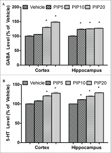
Figure 2. Characterization of electrophysiological effects of piperine. (A) Current voltage relations recorded prior and after application of 10 µM piperine to rNav1.4. Data from multiple paired experiments are included in the figure. Note the negative shift in half activation potential in the presence of piperine. (B) Steady state inactivation curves for rNav1.4 in the absence (wt) and presence of 10 µM piperine. No significant shift was observed. (C) Ensemble dose response curve for tonic rNav1.4 channel inhibition by piperine. The solid line is a fit via the Hill equation (IC50 10 µM ± 4 µM). (D) Effects of 10 µM piperine on rat Cav1.2, human LVA calcium channels and rat Nav1.4, (n = 4-5 per channel). Error bars reflect standard errors. The asterisk denotes that rNav1.4 block was statistically greater relative to the other channels (*p < 0.05).
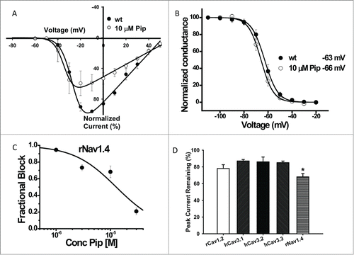
Table 5. Biophysical parameters of rNav1.4 sodium channels in the absence and the presence of piperine