Figures & data
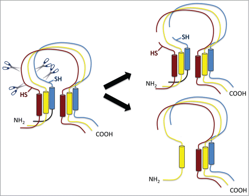
Figure 1. Schematic view of ENaC composed of 3 homologous subunits α (red), β (yellow) and gamma (blue) subunits with similar transmembrane topologies. The α and gamma subunits are cleaved at least twice in the extracellular domain near the N-terminal membrane-spanning segment (left). This divides the subunits into 2 segments. The shorter N-terminal part could stay within the channel structure (upper right) or separate (lower right). SH marks sites of engineered cysteines that affect channel function.