1,091
Views
17
CrossRef citations to date
0
Altmetric
Article Addendum

Cisplatin activates volume sensitive LRRC8 channel mediated currents in Xenopus oocytes

, , , & ORCID Icon
Pages 254-260 | Received 21 Dec 2016, Accepted 13 Jan 2017, Published online: 17 Feb 2017

Figures & data

Figure 1. Acute application of cisplatin and blasticidin does not affect currents mediated by 8A-VFP/8E-mCh or 8A-VFP/8D-mCh. (A) Typical current traces of 8A-VFP/8E-mCh expressing oocytes evoked by the “IV-pulse protocol” (see Materials and Methods) in control conditions and after perfusion of 1 mM cisplatin (top) or 0.25 mM blasticidin (bottom). (B) 8A-VFP/8E-mCh currents measured at 60 mV in presence of 1mM cisplatin or 0.25 mM blasticidin were normalized to the value measured before drug application (n = 5 for cisplatin; n = 3 for blasticidin). (C-D) Acute effect of cisplatin and blasticidin on 8A-VFP/8D-mCh. (C) Voltage clamp traces of oocytes expressing 8A-VFP/8D-mCh before and after perfusion of 1 mM cisplatin (top) or 0.25 mM blasticidin (bottom). (D) 8A-VFP/8D-mCh currents recorded at 60 mV were normalized to the value measured before drug application (n = 3). Error bars indicate SD.
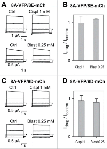
Figure 1. Acute application of cisplatin and blasticidin does not affect currents mediated by 8A-VFP/8E-mCh or 8A-VFP/8D-mCh. (A) Typical current traces of 8A-VFP/8E-mCh expressing oocytes evoked by the “IV-pulse protocol” (see Materials and Methods) in control conditions and after perfusion of 1 mM cisplatin (top) or 0.25 mM blasticidin (bottom). (B) 8A-VFP/8E-mCh currents measured at 60 mV in presence of 1mM cisplatin or 0.25 mM blasticidin were normalized to the value measured before drug application (n = 5 for cisplatin; n = 3 for blasticidin). (C-D) Acute effect of cisplatin and blasticidin on 8A-VFP/8D-mCh. (C) Voltage clamp traces of oocytes expressing 8A-VFP/8D-mCh before and after perfusion of 1 mM cisplatin (top) or 0.25 mM blasticidin (bottom). (D) 8A-VFP/8D-mCh currents recorded at 60 mV were normalized to the value measured before drug application (n = 3). Error bars indicate SD.

Figure 2. Cisplatin incubation strongly increases currents of oocytes expressing 8A-VFP/8E-mCh or 8A-VFP/8D-mCh. (A) Mean currents from un-injected or from 8A-VFP/8E-mCh injected oocytes measured before (t = 0; un-injected n = 29; 8A-VFP/8E-mCh n = 60) and after overnight incubation in “Maintaining” solution (Ctrl; un-injected n = 11; 8A-VFP/8E-mCh n = 29), 0.25 mM blasticidin (Blast 0.25; un-injected n = 4; 8A-VFP/8E-mCh n = 9), 0.5 mM cisplatin (Cispl 0.5; 8A-VFP/8E-mCh n = 7) or 1 mM cisplatin (Cispl 1; un-injected n = 14; 8A-VFP/8E-mCh n = 15; *****P < 10−9). The insets show 8A-VFP/8E-mCh traces from one oocyte before and after overnight incubation in 0.5 mM (left) or 1 mM cisplatin (right). (B) Bars represent currents of un-injected or 8A-VFP/8D-mCh injected oocytes recorded before (t = 0; un-injected n = 19; 8A-VFP/8D-mCh n = 39) and after overnight incubation in “Maintaining” solution (Ctrl; un-injected n = 8; 8A-VFP/8D-mCh n = 21), 0.15 mM cisplatin (Cispl 0.15; 8A-VFP/8D-mCh n = 6) or 1 mM cisplatin (Cispl 1; un-injected n = 11; 8A-VFP/8D-mCh n = 12; ***P < 0.001; ****P < 0.0001). The insets show typical 8A-VFP/8D-mCh traces from one oocyte before and after overnight incubation in 0.15 mM (left) or 1 mM cisplatin (right). Error bars indicate SD.
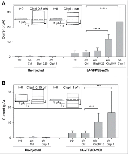
Figure 2. Cisplatin incubation strongly increases currents of oocytes expressing 8A-VFP/8E-mCh or 8A-VFP/8D-mCh. (A) Mean currents from un-injected or from 8A-VFP/8E-mCh injected oocytes measured before (t = 0; un-injected n = 29; 8A-VFP/8E-mCh n = 60) and after overnight incubation in “Maintaining” solution (Ctrl; un-injected n = 11; 8A-VFP/8E-mCh n = 29), 0.25 mM blasticidin (Blast 0.25; un-injected n = 4; 8A-VFP/8E-mCh n = 9), 0.5 mM cisplatin (Cispl 0.5; 8A-VFP/8E-mCh n = 7) or 1 mM cisplatin (Cispl 1; un-injected n = 14; 8A-VFP/8E-mCh n = 15; *****P < 10−9). The insets show 8A-VFP/8E-mCh traces from one oocyte before and after overnight incubation in 0.5 mM (left) or 1 mM cisplatin (right). (B) Bars represent currents of un-injected or 8A-VFP/8D-mCh injected oocytes recorded before (t = 0; un-injected n = 19; 8A-VFP/8D-mCh n = 39) and after overnight incubation in “Maintaining” solution (Ctrl; un-injected n = 8; 8A-VFP/8D-mCh n = 21), 0.15 mM cisplatin (Cispl 0.15; 8A-VFP/8D-mCh n = 6) or 1 mM cisplatin (Cispl 1; un-injected n = 11; 8A-VFP/8D-mCh n = 12; ***P < 0.001; ****P < 0.0001). The insets show typical 8A-VFP/8D-mCh traces from one oocyte before and after overnight incubation in 0.15 mM (left) or 1 mM cisplatin (right). Error bars indicate SD.

Figure 3. Cisplatin injection does not modify 8A-VFP/8E-mCh currents. Bars represent mean currents of un-injected or 8A-VFP/8E-mCh injected oocytes measured before (t = 0; un-injected n = 11; 8A-VFP/8E-mCh n = 15) and after overnight incubation in “Maintaining” solution. After recording currents in control conditions, some oocytes were just incubated (o/n Ctrl; un-injected n = 4; 8A-VFP/8E-mCh n = 8), others were injected with 50 nl of a 3 mM cisplatin solution and then incubated overnight (o/n inj Cispl 3; un-injected n = 7; 8A-VFP/8E-mCh n = 7; P > 0.05). Error bars indicate SD.
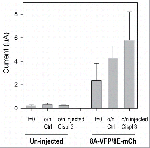
Figure 3. Cisplatin injection does not modify 8A-VFP/8E-mCh currents. Bars represent mean currents of un-injected or 8A-VFP/8E-mCh injected oocytes measured before (t = 0; un-injected n = 11; 8A-VFP/8E-mCh n = 15) and after overnight incubation in “Maintaining” solution. After recording currents in control conditions, some oocytes were just incubated (o/n Ctrl; un-injected n = 4; 8A-VFP/8E-mCh n = 8), others were injected with 50 nl of a 3 mM cisplatin solution and then incubated overnight (o/n inj Cispl 3; un-injected n = 7; 8A-VFP/8E-mCh n = 7; P > 0.05). Error bars indicate SD.

Figure 4. Cisplatin incubation does not alter currents of oocytes expressing WT 8A/8E. (A) Top: typical current traces measured in isotonic conditions of an 8A/8E expressing oocyte before and after overnight cisplatin incubation in the “maintaining” solution. Bottom: stimulation of the same oocyte after o/n incubation with hypotonic solution activates normal sized anion currents (left: current measured at 60 mV during hypotonic stimulation, right: voltage-clamp traces after hypotonic activation). (B) Mean values of 8A/8E currents in isotonic conditions before and after o/n cisplatin incubation (n = 7; P > 0.05). Error bars indicate SD.
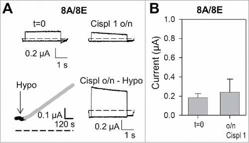
Figure 4. Cisplatin incubation does not alter currents of oocytes expressing WT 8A/8E. (A) Top: typical current traces measured in isotonic conditions of an 8A/8E expressing oocyte before and after overnight cisplatin incubation in the “maintaining” solution. Bottom: stimulation of the same oocyte after o/n incubation with hypotonic solution activates normal sized anion currents (left: current measured at 60 mV during hypotonic stimulation, right: voltage-clamp traces after hypotonic activation). (B) Mean values of 8A/8E currents in isotonic conditions before and after o/n cisplatin incubation (n = 7; P > 0.05). Error bars indicate SD.

Reprints and Corporate Permissions

Please note: Selecting permissions does not provide access to the full text of the article, please see our help page How do I view content?

To request a reprint or corporate permissions for this article, please click on the relevant link below:

Academic Permissions

Please note: Selecting permissions does not provide access to the full text of the article, please see our help page How do I view content?

Obtain permissions instantly via Rightslink by clicking on the button below:

If you are unable to obtain permissions via Rightslink, please complete and submit this Permissions form. For more information, please visit our Permissions help page.