617
Views
0
CrossRef citations to date
0
Altmetric
Review Article

Ionotropic purinergic receptor 7 (P2X7) channel structure and pharmacology provides insight regarding non-nucleotide agonism

& ORCID Icon
Article: 2355150 | Received 01 Apr 2024, Accepted 10 May 2024, Published online: 19 May 2024

Figures & data

Figure 1. Functionally important binding sites aligned on full-length P2X7 structure. (a) full-length structures of rat P2X7 in the putative closed (Apo, 6U9V) and open (ATP, 6U9W) states. (b,c) top-down (top) and side-on (bottom) views of the three, inter-subunit, ATP binding sites (B, ATP), and single orthosteric binding sites (B) and allosteric binding sites (C). (d) side-on view of a single P2X7 channel subunit (6U9V) to highlight locations of relevant binding sites and functionally important features.
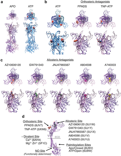
Four sets of images of high-resolution crystal structures of the P2X7 ion channel with top-down and side-on views showing the channel in nine different ligand-bound states.
Figure 1. Functionally important binding sites aligned on full-length P2X7 structure. (a) full-length structures of rat P2X7 in the putative closed (Apo, 6U9V) and open (ATP, 6U9W) states. (b,c) top-down (top) and side-on (bottom) views of the three, inter-subunit, ATP binding sites (B, ATP), and single orthosteric binding sites (B) and allosteric binding sites (C). (d) side-on view of a single P2X7 channel subunit (6U9V) to highlight locations of relevant binding sites and functionally important features.

Table 1. P2X7 agonists, antagonists, and modulators: effects on function and/or expression.

Table 2. P2X7-selective and nonselective antagonist co-crystals.

Table 3. P2X7 inhibitors in clinical trials.

Data availability statement

Data sharing is not applicable to this article as no new data were created in this study.