Figures & data
Figure 1. WT and BLT2 knockout mice show a similar trend in weight gain in response to control or high fat diet. Weekly weight measurements in non-sedated animals for 5 weeks (mean ± SEM), n = 5 per group **p < 0.01 2-Way ANOVA.
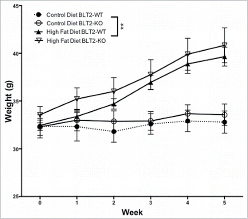
Table 1. Metabolic and biochemical characteristics after 5 weeks of controlled diet (average ± SEM), n = 5 per group, * or § p < 0.05, ** or ¶¶ or §§ p < 0.01 non parametric one-way ANOVA with Tukey's post hoc test.
Figure 2. BLT2 knockout mice show an abrogated glucose response, sign of spontaneous T2D. (A) Glucose tolerance test after 5 weeks of controlled diet in WT and knockout mice, 6 measurements were taken after 1 g/kg glucose IP injection (mean ± SEM). n = 5 animals per group **p < 0.01 2-Way ANOVA. (B) Area under the glucose tolerance test curves (mean ± SEM). n = 5 animals per group *p < 0.05 **p < 0.01, non-parametric One-Way ANOVA with Tukey's post hoc test.
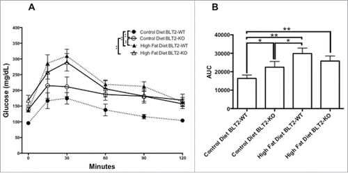
Figure 3. The skin from BLT2 KO and T2D mice present atrophic epidermis and reduced TEER. (A) Microphotographs MT stains at 20x magnification and representative insets at double magnification, (B) semi-quantification of epidermal thickness and (C) Ex vivo TEER. The data shown represent the mean ± SEM, n = 5 animals per group ***p < 0.01, non-parametric One-Way ANOVA with Tukey's post hoc test.
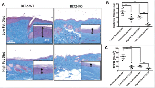
Figure 4. The skin from BLT2 knockout mice exhibits increased levels of inflammatory, matrix degradation and differentiation markers compared with tissue from T2D animals. Q-PCR for (A) Il1b, (B) Cxcl2, (C) Mmp9, (D) Flg, (E) Lor and (F) Krt10, relative to actb in skin from WT and BLT2 knockout mice under control or high fat diet for 5 weeks. Data represent the mean ± SEM of n = 5 per group, *p < 0.05 **p < 0.01 non-parametric One-Way ANOVA with Tukey's post hoc test.
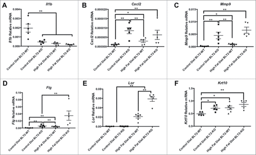
Figure 5. High glucose levels disorganize the actin structure disrupting the monolayer and reducing the TEER in HaCaT cells, an alteration that is prevented by BLT2 expression. (A) Representative confocal microphotographs of actin fluorescence stain (Phalloidin-FITC) in HaCaT cells after 48 h under low (5 mM) and high (25 mM) glucose levels. In blue: nucleus stain by DAPI. n = 3 independent experiments. (B) Transepithelial resistance (TEER) assay in high or low glucose levels after 48 h. Data represent the mean ± SEM of n = 3 independent experiments *p < 0.05 non parametric One-Way ANOVA with Tukey's post hoc test.
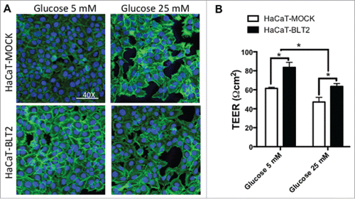
Figure 6. High glucose reduces wound healing capacity, an effect that is attenuated in cells that express BLT2. (A) Representative micrographs of wound healing assays in HaCaT cells, for 48 h under low (5 mM) and high (25 mM) glucose, (B) typical kinetic curve of migration and wound closure. Data represent the mean ± SEM, n = 6 independent experiments **p < 0.01 2-Way ANOVA.
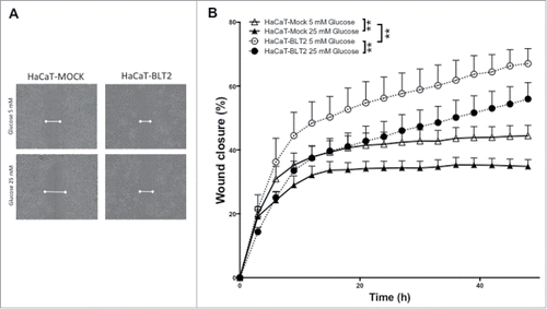
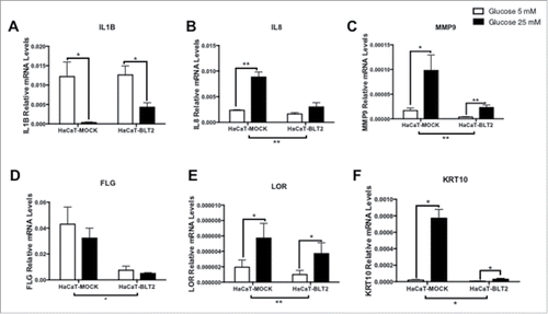
Figure 7. Cells that do not express BLT2 present altered levels of inflammatory, matrix degradation and differentiation markers. High glucose exerts a more severe effect on these cells. Q-PCR for IL1B, IL8, MMP9, FLG, LOR and KRT10 relative to ACTB, in HaCaT Mock and BLT2 after 48 h of low (5 mM) or high (25 mM) glucose. Data represent the mean ± SEM, n = 3 independent experiments run in duplicate *p < 0.05 **p < 0.01 non-parametric One-Way ANOVA with Tukey's post hoc test.