Figures & data
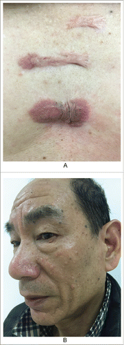
Figure 1. the nature history of keloid. A. From outside to inside: the bright red uplift patches/nodules, growing rapidly; inner ring static relatively,with dark color; then the middle shows atrophy.(from Bolognia JL,Jorizzo JL,Schaffer JV .Dermatology (3rd Edition). Amsterdam(NL):Elsevier;2012.p1622.Fig.98.2B) B. The atrophic scar in the breast with the re-proliferation at the edge.
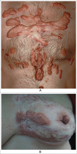
Figure 2. We emphasize repeated inflammation stimulates to the granulation tissue hyperplasia in the area without suture, so leaving scars after healing. (from Bolognia JL,Jorizzo JL,Schaffer JV .Dermatology (3rd Edition). Amsterdam(NL):Elsevier;2012.p2315.Fig.141.4)
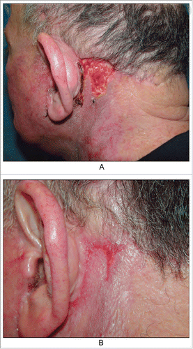
Figure 3. Keloids secondary to severe acne. A. Obvious cord-like hyperplasia on the back; B. Nodules and cysts on the submandibular neck, old scars/keloids on the neck and neck side; C. The contents of sebaceous glands were squeezed out due to the high injection pressure, when we injected the hormone into scar nodules.
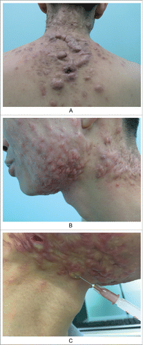
Figure 4. A keloid after acne in cheek. A. Before; B. After six months of treatment, scars flattened. But epidermoid cysts were found around the scar. C. A epidermoid cyst which is already scarring.
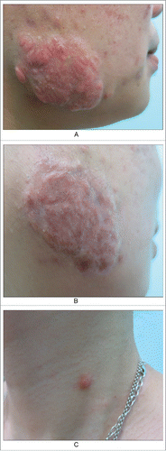
Figure 5. The patient with keloids in the chest. A. Before B. After four months treament of glucocorticoid and 5-fluorouracil, leaving a lot of blackheads and epidermoid cysts.
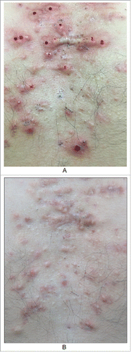
Figure 6. Patient showing keloid and sebaceous gland hyperlplasia. A. The keloids in his chest for 20 years. B. Multiple sebaceous gland hyperplasia in his face.