Figures & data
Figure 1. (A) Diagram of the experimental procedure allowing the implantation of the developing pancreatic rudiments in the AC of the eye at E13.0. (B) Contraption allowing the stabilization of the mouse.
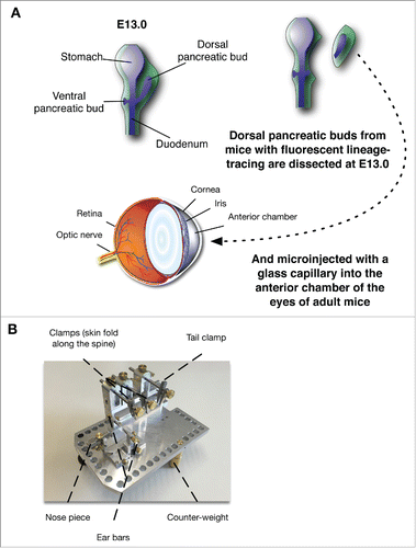
Figure 2. Brightfield images of the growing pancreatic anlages growing in the AC of the eye 4 d (A) and 7 d (B) after implantation. The dotted lines delimit the developing rudiment.
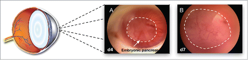
Figure 3. Brightfield and fluorescent images of a Pdx1-Cre; YFP pancreatic rudiment at E13 right before implantation (A and A’ respectively); 4 d (B and B’ respectively), and 7 d after implantation (EDF images in C and C’ respectively). (D) Immunofluorescence for Insulin (blue), Glucagon (red) and CP-A (green) on sections from rudiments implanted for 7 d. (E) Immunofluorescence for E-Cadherin (red), the YFP reporter with a DAPI counterstain in a cross-section of the eye with a rudiment from a Pdx-Cre; YFP mouse. (F) Hematoxylin and Eosin stain, (G) Eosin stain and (H) immunofluorescence for Vimentin (red), YFP (green) with a DAPI counterstain on sections from Pdx-Cre; YFP rudiments implanted for 7 d Scale bars=50 μm.
Figure 4. (A) Diagram representing the partial reduction in mesenchyme before implantation. Brightfield and fluorescent images of a Pdx1-Cre; YFP pancreatic rudiment at E13 with reduced mesenchyme right before implantation (B and B’ respectively); 2 d (C and C’ respectively), 12 d after implantation (D and D’ respectively), 18 d after implantation (E and E’ respectively) and as the eye has been removed at the end of the experiment at 18 d after implantation (F and F’ respectively). (G) Immunofluorescence for CP-A (red), and YFP (green) on sections from the rudiments implanted for 18 d H. Immunofluorescence for Insulin (blue), Glucagon (red) and YFP (green) on sections from the rudiments implanted for 18 d (I) Immunofluorescence for Sox9 (red) and YFP (green) with a DAPI nuclear counterstain (blue) on sections from the rudiments implanted for 18 d Scale bars = 50 μm.
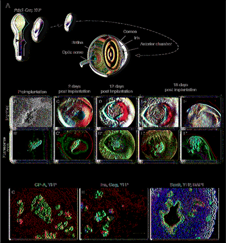
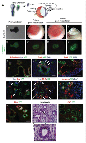
Figure 5. (A) Diagram representing the near-complete reduction in mesenchyme before implantation. Brightfield and fluorescent images of a Pdx1-Cre; YFP pancreatic rudiment at E13 with almost no mesenchyme right before implantation (B and B’ respectively); 5 d (C and C’ respectively) and 7 d after implantation (D and D’ respectively) and as the eye has been removed at the end of the experiment at 7 d after implantation (E and E’ respectively). (F) Immunofluorescence on sections from the rudiments implanted for 7 d for E-Cadherin (red), Insulin (blue) and YFP (green) (F); Pdx1 (red) and YFP (green) with a DAPI nuclear counterstain (blue; G); Sox9 (red) and YFP (green) with a DAPI nuclear counterstain (blue; H); Insulin (blue), Glucagon (red) and YFP (green; I); Insulin (blue), CP-A (red) and YFP (green; J); Amylase (red) and YFP (green) with a DAPI nuclear counterstain (blue; K); Insulin (blue), Glucagon (red) and YFP (green; I); DBA (red) and YFP (green; L); Hematoxylin staining (M with a magnification below) and vWF (red) and YFP (green; N). Scale bars = 50 μm. Scale bars = 50 μm.