Figures & data
Comparison of the Length of Meiotic Spindle Plate of In Vivo and In Vitro Matured Oocytes Derived from Naturally Cycling or Gonadotropin Stimulated Mice (5 Replicates)*
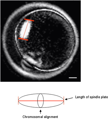
Figure 1 Measurement for the length of spindle plate of mouse oocyte using the PolScope imaging system. The length of spindle plate was measured by crossing the chromosomal alignment (arrow). Scale bar indicates 10 μm.
Comparison of Meiotic Spindle Organization and Chromosomal Alignment of In Vivo and In Vitro Matured Oocytes Derived from Naturally Cycling or Gonadotropin Stimulated mice (10 Replicates)*
Figure 2 Morphology of meiotic spindle organization and chromosome alignment in normal and abnormal mouse oocytes. (A) Oocyte with normal meiotic spindle organization (green); (a) The same oocyte with normal chromosome alignment (blue); (B) Oocyte with abnormal meiotic spindle organization (green); (b) The same oocyte with abnormal chromosome alignment (blue). Scale bar indicates 20 μm.
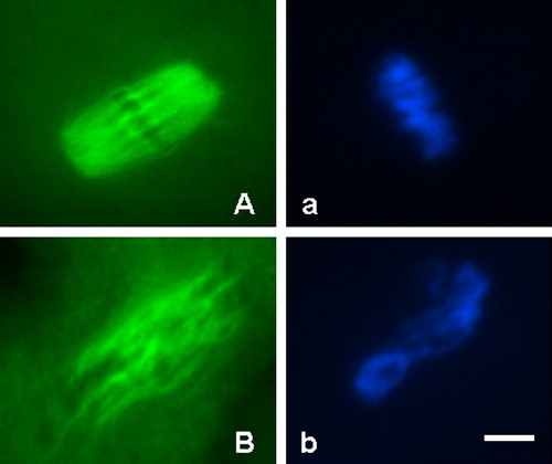
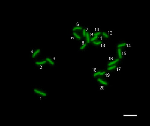
Figure 3 Normal (haploidy) chromosomes in parthenogenetically activated mouse oocyte. The oocyte with 20 chromosomes. Scale bar indicates 20 μm.
Ploidy Analysis of In Vivo and In Vitro Matured Oocytes Derived from Naturally Cycling or Gonadotropin Stimulated Mice (6 Replicates)*