
Figures & data
Table 1. Phospholipids (PL) and triacylglycerols (TAG) identified via MALDI(+)-MS in the supplements, in the oocytes, and in the denuded oocytes.
Figure 1. MALDI-MS lipid profiles of serum used as supplements for oocytes IVM, before solubilization to culture medium. Spectra were obtained in the positive mode using 2,5-Dihydroxybenzoic acid (DHB) as matrix. Analyses were performed directly on fetal bovine serum - FBS (A) and synthetic serum substitute – SSS (B).
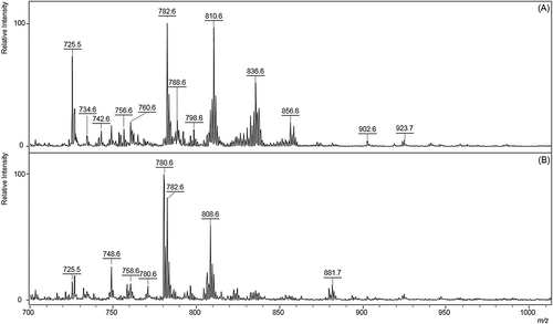
Figure 2. MALDI-MS lipid profiles of pooled cumulus cells from immature and mature oocytes supplemented with sera during 24 h of IVM. (A) iCC: immature cumulus cells; (B) FBS-CC: cumulus cells obtained from FBS-supplemented oocytes after IVM; (C) SSS-CC: cumulus cells obtained from SSS-supplemented oocytes after IVM. FBS: fetal bovine serum; SSS: synthetic serum substitute; IVM: in vitro maturation. Spectra were obtained in the positive mode using 2,5-Dihydroxybenzoic acid (DHB) as matrix.
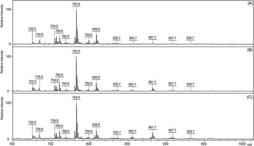
Figure 3. Three-dimensional PLS-DA score plot of samples from immature cumulus cells (iCC), FBS-treated cumulus cells (FBS-CC), and SSS-treated cumulus cells (SSS-CC) during IVM. FBS: fetal bovine serum; SSS: synthetic serum substitute; IVM: in vitro maturation. A clear separation between three clusters was achieved. See Supplemental Figure S2 for related variable importance in projection (VIP) plot of the ions.
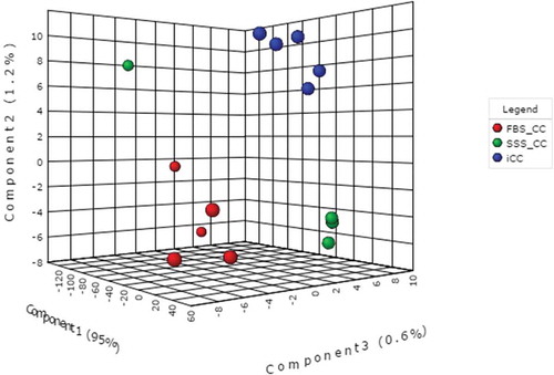
Figure 4. MALDI-MS lipid profiles of immature oocytes (IO) and oocytes supplemented with sera during IVM in the presence (COC) or absence (DO) of cumulus cells. Spectra were obtained in the positive mode using 2,5-dihydroxybenzoic acid (DHB) as matrix. Analyses were performed directly on pools of 5 mechanically denuded oocytes from 15 replicates. (A) IO; (B) FBS-COC; (C) SSS-COC; (D) FBS-DO; and (E) SSS-DO. IVM: in vitro maturation; COC: cumulus-oocyte complex; DO: denuded oocyte; FBS: fetal bovine serum; SSS: synthetic serum substitute; CC: cumulus cells.
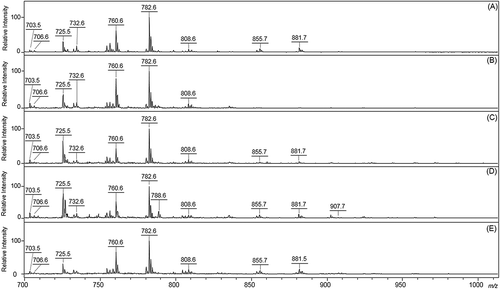
Figure 5. Three-dimensional PLS-DA score plot of samples from FBS-treated denuded oocytes (FBS-DO) and FBS-treated cumulus-oocyte complexes (FBS-COC). A good separation between two clusters was achieved. See Supplemental Figure S4 for related variable importance in projection (VIP) plot of the ions. FBS: fetal bovine serum; SSS: synthetic serum substitute.
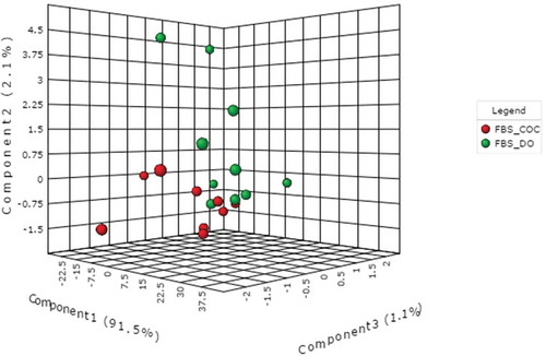
Figure 6. Three-dimensional PLS-DA score plot of samples from SSS-treated denuded oocytes (SSS-DO) and SSS-treated cumulus-oocyte complexes (SSS-COC). A clear separation between two clusters was achieved. See Supplemental Figure S6 for related variable importance in projection (VIP) plots reporting the ions responsible by clusters separation. FBS: fetal bovine serum; SSS: synthetic serum substitute.
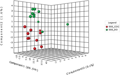
Table 2. Embryonic development of bovine denuded oocytes (DO) or cumulus-oocyte-complexes (COC) matured in vitro in TCM-199 medium supplemented with fetal bovine serum (FBS) or synthetic serum substitute (SSS).