Figures & data
Figure 1. Levels of testosterone (A), follicular stimulating hormone (FSH) (B) and luteinizing hormone (LH) (C) in the serum of all experimental groups of rats. All measurements were done by ELISA. Values are expressed as Mean ± SD for 12 rats in each group. Values were considered significantly different at p < 0.05. aSignificantly different when compared to the control group, bsignificantly different when compared to resveratrol (RES)-treated group and csignificantly when compared to cisplatin (Cis)-treated group.
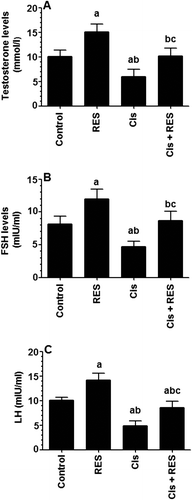
Table 1. Testes and final body weights in all groups of rats.
Table 2. Characterization of epididymal sperm morphology in groups of rat.
Figure 2. Average sperm count and motility in all groups of rats. Sperm analysis was done using the right cauda epididymis. The total number of sperm and sperm motility were calculated using a hemocytometer. For sperm morphological analysis, sperm structures were visualized under a light microscope after adding a drop of Eosin to sperm suspension. Values were considered significantly different at p < 0.05. aSignificantly different when compared to the control group, bsignificantly different when compared to resveratrol (RES)-treated group and csignificantly when compared to cisplatin (Cis)-treated group.
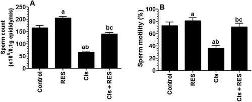
Figure 3. Levels of reduced glutathione (GSH), glutathione disulfide (GSSG) (A), tumor necrosis factor-α (TNF-α) (B) and interleukin-6 (IL-6) (C) in the testis homogenates of all groups of rats. Levels of GSH and GSSG were measured by colorimetric-based kits were as levels of TNF-α and interleukin-6 (IL-6) were measured by ELISA. Values are expressed as Mean ± SD for 6 rats in each group. Values were considered significantly different at p < 0.05. aSignificantly different when compared to the control group, bsignificantly different when compared to resveratrol (RES)-treated group and csignificantly when compared to cisplatin (Cis)-treated group.
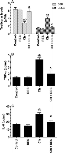
Figure 4. Protein levels of glutathione reductase (GRx), superoxide dismutase (SOD), catalase (CAT) and 4-hydroxynonenal (4HNE) in the testis of all groups of rats as relatively expressed to the reference protein, GAPDH. For western blotting, equal volumes of protein sample (60 µg protein/well) were separated by an SDS electrophoresis, transferred to nitrocellulose membrane and then incubated with the primary and corresponding horseradish peroxidase-conjugated antibodies. Antigen-antibody complexes were then visualized using a Pierce ECL kit and images were quantified using Image J software. Values are expressed as Mean ± SD for 6 rats in each group. Values were considered significantly different at p < 0.05. aSignificantly different when compared to the control group (lane 1), bsignificantly different when compared to resveratrol (RES)-treated group (Lane 2) and csignificantly when compared to cisplatin (Cis)-treated group (Lane 3). Lane 4: Cis +RES-treated group.
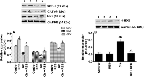
Figure 5. Protein levels of components of intrinsic (mitochondrial) apoptotic pathway in the testis of all groups of rats as relatively expressed to the reference protein, GAPDH. For western blotting, equal volumes of protein sample (60µg protein/well) were separated by an SDS electrophoresis, transferred to nitrocellulose membrane and then incubated with the primary and corresponding horseradish peroxidase-conjugated antibodies. Antigen-antibody complexes were then visualized using a Pierce ECL kit and images were quantified using Image J software. Values are expressed as Mean ± SD for 6 rats in each group as compared to corresponding GAPDH levels. Values were considered significantly different at p < 0.05. aSignificantly different when compared to control the group (lane 1), bsignificantly different when compared to resveratrol (RES)-treated group (Lane 2) and csignificantly when compared to cisplatin (Cis)-treated group (Lane 3). Lane 4: Cis+ RES-treated group. L: Ladder.
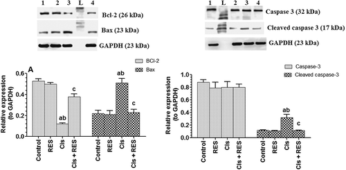
Figure 6. Protein levels of phospho-p42 and phosphor-p44 mitogen-activated protein kinases (ERK1/2) and phospho-P45 and phosphor-P54 stress-activated protein kinase/Jun-amino-terminal kinase (SAPK/JNK) pathway in the testis of all groups of rats as expressed to the reference protein, GAPDH. For western blotting, equal volumes of protein sample (60µg protein/well) were separated by an SDS electrophoresis, transferred to nitrocellulose membrane and then incubated with the primary and corresponding horseradish peroxidase-conjugated antibodies. Antigen-antibody complexes were then visualized using a Pierce ECL kit and images were quantified using Image J software. Values are expressed as Mean ± SD for 6 rats in each group. Values were considered significantly different at p < 0.05. aSignificantly different when compared to control group (lane 1), bsignificantly different when compared to resveratrol (RES)-treated group (Lane 2) and csignificantly when compared to cisplatin (Cis)-treated group (Lane 3). Lane 4: Cis + RES--treated group.
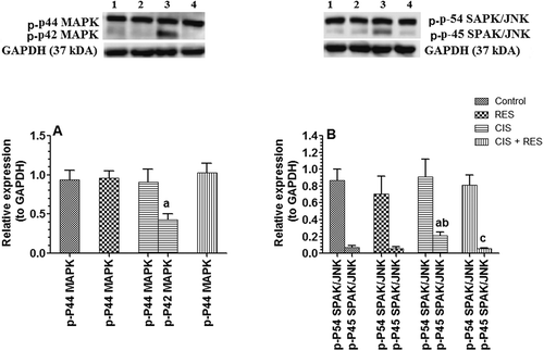
Figure 7. Protein levels of P53 in the testis of all groups of rats. Values are expressed as Mean ± SD for 6 rats in each group as relatively expressed to the reference protein, GAPDH. For western blotting, equal volumes of protein sample (60µg protein/well) were separated by an SDS electrophoresis, transferred to nitrocellulose membrane and then incubated with the primary and corresponding horseradish peroxidase-conjugated antibodies. Antigen-antibody complexes were then visualized using a Pierce ECL kit and images were quantified using Image J software. Values were considered significantly different at p < 0.05. aSignificantly different when compared to the control group (lane 1), bsignificantly different when compared to resveratrol (RES)-treated group (Lane 2) and csignificantly when compared to cisplatin (Cis)-treated group (Lane 3). Lane 4: Cis +RES–treated group.
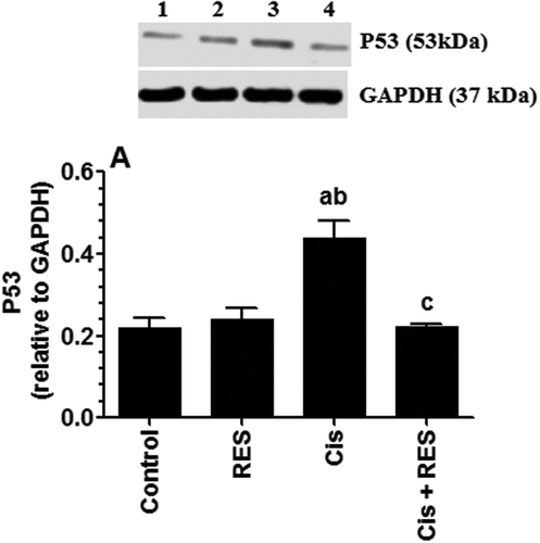
Figure 8. Protein levels of total and cleaved caspase-12 as well calpain-1 in the testis of all groups of rats as compared to corresponding GAPDH levels as relatively expressed to the reference protein, GAPDH. Values are expressed as Mean ± SD for 6 rats in each group. For western blotting, equal volumes of protein sample (60µg protein/well) were separated by an SDS electrophoresis, transferred to nitrocellulose membrane and then incubated with the primary and corresponding horseradish peroxidase-conjugated antibodies. Antigen-antibody complexes were then visualized using a Pierce ECL kit and images were quantified using Image J software. Values were considered significantly different at p < 0.05. aSignificantly different when compared to the control group (lane 1), bsignificantly different when compared to resveratrol (RES)-treated group (Lane 2) and csignificantly when compared to cisplatin (Cis)-treated group (Lane 3). Lane 4: Cis + RES--treated group.
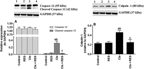
Figure 9. Protein of Akt, phosphor-Akt and Bad pathway in the testis of all groups of rats as expressed to the reference protein, GAPDH.Values are expressed as Mean ± SD for 6 rats in each group. For western blotting, equal volumes of protein sample (60 µg protein/well) were separated by an SDS electrophoresis, transferred to nitrocellulose membrane and then incubated with the primary and corresponding horseradish peroxidase-conjugated antibodies. Antigen-antibody complexes were then visualized using a Pierce ECL kit and images were quantified using Image J software. Values were considered significantly different at p < 0.05. aSignificantly different when compared to the control group (Lane 1), bsignificantly different when compared to resveratrol (RES)-treated group (Lane 2) and csignificantly when compared to cisplatin (Cis)-treated group (Lane 3). Lane 4: Cis +RES–treated group. L: Ladder.
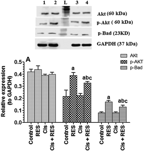
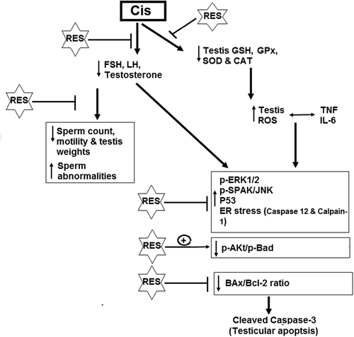
Figure 10. Schematic diagram of the mechanism of action of resveratrol against cisplatin-induced reproductive dysfunction.