
Figures & data
Figure 1. In vitro characterization of fusion proteins
(a) schematic representation of membrane-anchored LIGHT and cognate receptors HVEM and LTβR. (b) schematic representation of five LIGHT-based fusion proteins, with respective size exclusion chromatography profiles. The homotrimeric form of LIGHT expressed as a single polypeptide chain was fused to the C-terminus of (from left to right): the heavy chain of F8 in IgG format, the light chain of F8 in IgG format, the F8 in scFv-Fc format, the F8 in diabody format and the F8 in single-chain diabody format (F8-LIGHT). Detailed linear structure of F8-LIGHT is highlighted. (c,d) biochemical characterization of F8-LIGHT including SDS-PAGE of F8-LIGHT under non-reducing (NR) and reducing (R) conditions (c) and mass spectrometry profile of PNGase F-treated F8-LIGHT (calculated mass = 101791 Da) (d). (e) binding of titrated concentrations of F8-LIGHT and positive control SIP(F8) to immobilized target antigen EDA, measured by ELISA. (f) activity of F8-LIGHT, measured by a cytotoxicity assay on HT-29 cells in the presence of human Interferon gamma (hIFNγ). Reported concentrations are based on the molecular weight of the LIGHT part of the molecule alone. 7-AAD positive dead cells were detected by Flow Cytometry. Column represent means ± SEM, n = 3 per experimental group, ns = non-significant, * = p < .05, ** = p < .01, *** p = < 0.001, **** = p < .0001 (unpaired t-test).
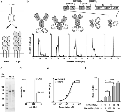
Figure 2. Tumor targeting of F8-LIGHT in vivo.
Biodistribution experiment in 129/Sv mice bearing F9 tumor. Radio-iodinated F8-LIGHT and KSF-LIGHT (used as negative control) were injected in the lateral tail vein. Accumulation of the fusion proteins in tumor and healthy organs after 24 hours was calculated as percentage of injected dose per gram of tissue (% ID/g). Column represent means ± SEM, n = 4 per experimental group, * = p < .05, ** = p < .01, *** p = <0.001, **** = p < .0001 (unpaired t-test).
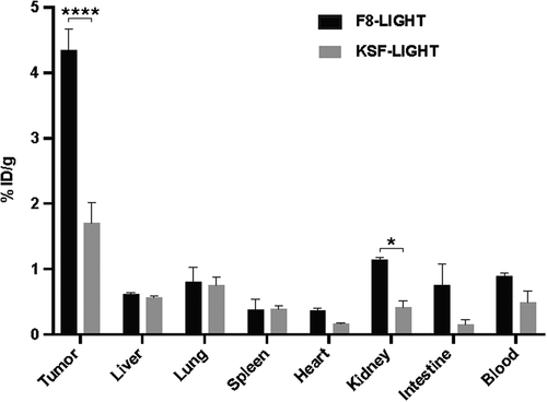
Figure 3. Therapy experiments
(a) Therapy experiment in BALB/c mice bearing established CT26 tumor. 100 μg of F8-LIGHT or KSF-LIGHT were administered intravenously every other day, as indicated by the black arrows. (b) as in (a), but mice received 300 μg of F8-LIGHT. (c) Therapy experiment in BALB/c mice bearing established CT26 tumor. 300 μg of F8-LIGHT or 150 μg of F8 (black arrows), 200 μg of anti-PD-1 (gray arrows) or a combination of F8-LIGHT and anti-PD-1 were administered as depicted in the figure. (d) Therapy experiment in BALB/c mice bearing established WEHI-164 tumor. 300 μg of F8-LIGHT (black arrows), 200 μg of anti-PD-1 (gray arrows) or a combination of the two were administered as depicted in the figure. Data represent means ± SEM, n = 5 mice per experimental group. * = p < .05, ** = p < .01, *** p = <0.001, **** = p < .0001 (regular two-way ANOVA test with Bonferroni posttest correction).
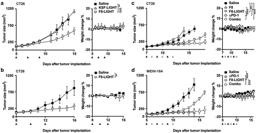
Figure 4. Analysis of immune infiltrate
Analysis of tumors and tumor-draining lymph nodes (TDLN) of BALB/c mice bearing WEHI-164 tumors, 48 hours after the third administration of F8-LIGHT or saline. (a) lymphocytes infiltration in tumors and composition of TDLN. CD3 = CD3+ lymphocytes, CD8 = CD8 + T cells, CD4 = CD4 + T cells, Treg = CD4+ regulatory T cells, NKT = Natural Killer T cells, NK = Natural Killer cells, APC = Antigen Presenting cells and MHCII+NK = MHC class II+ NK cells. (b) total NK1.1-positive and MHC class II-positive lymphocytes infiltrating tumors. (c) phenotype of CD8 + T cells and of AH1-specific CD8 + T cells in tumors and TDLN, based on expression of the markers CD44 and CD62L. Teff = effector T cells, Tnaive = naïve T cells, Tcm = central memory T cells. (d) AH1-specific CD8 + T cells in tumors and TDLN. (e) CD8 + T cells:CD4 + T cells ratio in tumor and TDLN. Data represent individual mice, with means ± SEM, n = 3 mice per experimental group. * = p < .05, ** = p < .01, *** p = <0.001 (unpaired t-test).
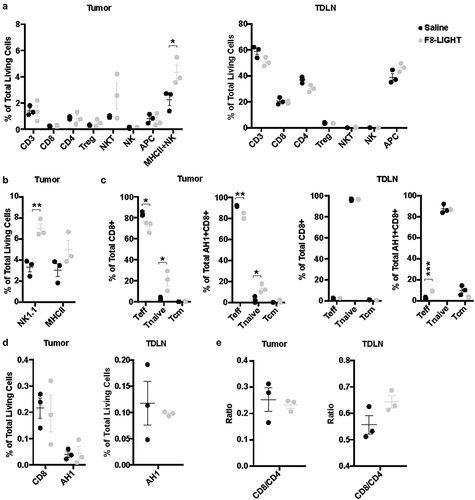
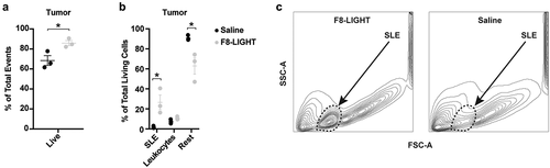
Figure 5. Analysis of tumor composition
WEHI-164 tumors analysis 48 hours after the third administration of F8-LIGHT or saline (a) fraction of living cells among total events recorded. (b) composition of living cells in the tumor. “Leukocytes” represent the sum of all T cells, NK cells, Antigen Presenting cells and Granulocytes, “Rest” represent the remaining living cells, after subtracting SLE and Leukocytes from the total number of living cells. SLE = “Small Living Events” C, representative analysis of tumor cells suspensions from single mice treated with F8-LIGHT or saline. SSC-A = side scatter area, FSC-A = forward scatter area. Data represent individual mice, with means ± SEM, n = 3 mice per experimental group. * = p < .05 (unpaired t-test).
Supplemental material
Supplemental Material
Download MS Word (3.2 MB)Data availability statement
Data and material are presented in the main text and supplementary information, raw data are available from the corresponding author on reasonable request.