4,500
Views
17
CrossRef citations to date
0
Altmetric
Report

Brain pharmacokinetics of anti-transferrin receptor antibody affinity variants in rats determined using microdialysis

ORCID Icon, , , , , & ORCID Icon show all
Article: 1874121 | Received 02 Nov 2020, Accepted 06 Jan 2021, Published online: 26 Jan 2021

Figures & data

Table 1. Non-compartmental analysis of plasma and brain PK profiles of anti-TfR antibody affinity variants in rats

Figure 1. In vivo plasma PK profiles of anti-TfR antibody affinity variants following 10 mg/kg systemic administration in rats. Open triangle: OX26-5 (KD = 5 nM), open square: OX26-76 (KD = 76 nM), closed circle: OX26-108 (KD = 108 nM), cross: OX26-174 (KD = 174 nM). Error bar: standard deviation
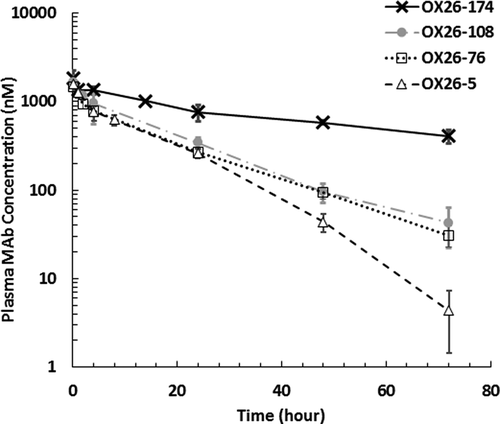
Figure 1. In vivo plasma PK profiles of anti-TfR antibody affinity variants following 10 mg/kg systemic administration in rats. Open triangle: OX26-5 (KD = 5 nM), open square: OX26-76 (KD = 76 nM), closed circle: OX26-108 (KD = 108 nM), cross: OX26-174 (KD = 174 nM). Error bar: standard deviation

Figure 2. In vivo PK of anti-TfR antibody affinity variants in different regions of the rat brain following 10 mg/kg systemic administration. (a)-(c) Free OX26 variant concentrations in: (A) cerebrospinal fluid (CSF) at the lateral ventricle (LV), (b) CSF at the cisterna magna (CM), and (C) brain interstitial fluid (ISF) at the striatum (ST). (d) Total OX26 variant concentrations in the whole brain homogenate. (e)-(g) CNS-to-plasma concentration ratios for unbound OX26 variants over the time. (E) CSFLV-to-plasma, (f) CSFCM-over-plasma, (G) ISF-to-plasma. (h) Brian-to-plasma concentration ratios for total OX26 variants. Closed triangle: OX26-5 (KD = 5 nM), open circle: OX26-76 (KD = 76 nM), mark: OX26-108 (KD = 108 nM), open square: OX26-174 (KD = 174 nM); LV: lateral ventricle, CM: cisterna magna, ST: striatum, error bar: standard deviation
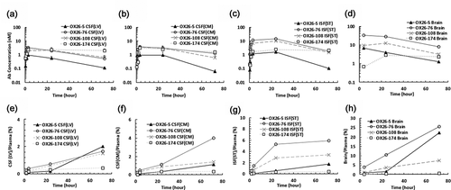
Figure 2. In vivo PK of anti-TfR antibody affinity variants in different regions of the rat brain following 10 mg/kg systemic administration. (a)-(c) Free OX26 variant concentrations in: (A) cerebrospinal fluid (CSF) at the lateral ventricle (LV), (b) CSF at the cisterna magna (CM), and (C) brain interstitial fluid (ISF) at the striatum (ST). (d) Total OX26 variant concentrations in the whole brain homogenate. (e)-(g) CNS-to-plasma concentration ratios for unbound OX26 variants over the time. (E) CSFLV-to-plasma, (f) CSFCM-over-plasma, (G) ISF-to-plasma. (h) Brian-to-plasma concentration ratios for total OX26 variants. Closed triangle: OX26-5 (KD = 5 nM), open circle: OX26-76 (KD = 76 nM), mark: OX26-108 (KD = 108 nM), open square: OX26-174 (KD = 174 nM); LV: lateral ventricle, CM: cisterna magna, ST: striatum, error bar: standard deviation

Figure 3. The influence of binding kinetic parameters to antibody exposure in the central nervous system. (a) The relationship between the AUC in different regions of the brain after 10 mg/kg i.v. administration of OX26 variants in rats and dissociation constants (KD) for TfR. A bell-shaped relationship was observed in the ISF and brain, but not in the CSF compartments. (b) The relationship between the AUC and dissociation rate constants (koff). AUC was computed from time 0 to the last time point (72 h). Open square: AUCbrain, closed circle: AUCISF(ST), closed triangle: AUCCSF(CM), and open diamond: AUCCSF(LV). AUC: area under the concentration curve, ISF: interstitial fluid, CSF: cerebrospinal fluid, ST: striatum, CM: cisterna magna, LV: lateral ventricle
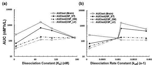
Figure 3. The influence of binding kinetic parameters to antibody exposure in the central nervous system. (a) The relationship between the AUC in different regions of the brain after 10 mg/kg i.v. administration of OX26 variants in rats and dissociation constants (KD) for TfR. A bell-shaped relationship was observed in the ISF and brain, but not in the CSF compartments. (b) The relationship between the AUC and dissociation rate constants (koff). AUC was computed from time 0 to the last time point (72 h). Open square: AUCbrain, closed circle: AUCISF(ST), closed triangle: AUCCSF(CM), and open diamond: AUCCSF(LV). AUC: area under the concentration curve, ISF: interstitial fluid, CSF: cerebrospinal fluid, ST: striatum, CM: cisterna magna, LV: lateral ventricle

Figure 4. In vivo PK of anti-TfR antibody affinity variants (OX26-5, OX26-76, OX26-108, and OX26-174) in rat plasma, brain interstitial fluid, whole-brain homogenate, isolated brain capillaries, and postvascular supernatant. The animals received 10 mg/kg i.v. dose of antibodies via the tail vein. Upper left: OX26-5 (KD = 5 nM), upper right: OX26-76 (KD = 76 nM), bottom left: OX26-108 (KD = 108 nM), bottom right: OX26-174 (KD = 174 nM). Open circle: plasma, open square: brain ISF, closed diamond: whole brain, closed square: postvascular supernatant, and closed triangle: isolated brain capillaries. The error bar presents the standard deviation
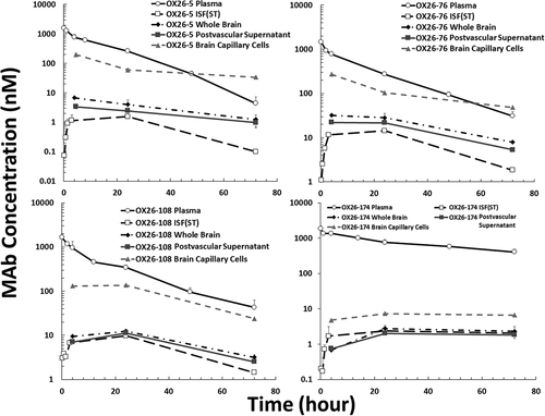
Figure 4. In vivo PK of anti-TfR antibody affinity variants (OX26-5, OX26-76, OX26-108, and OX26-174) in rat plasma, brain interstitial fluid, whole-brain homogenate, isolated brain capillaries, and postvascular supernatant. The animals received 10 mg/kg i.v. dose of antibodies via the tail vein. Upper left: OX26-5 (KD = 5 nM), upper right: OX26-76 (KD = 76 nM), bottom left: OX26-108 (KD = 108 nM), bottom right: OX26-174 (KD = 174 nM). Open circle: plasma, open square: brain ISF, closed diamond: whole brain, closed square: postvascular supernatant, and closed triangle: isolated brain capillaries. The error bar presents the standard deviation

Figure 5. The Push-Pull system for large-pore microdialysis. The system requires both a syringe pump and a peristaltic pump to maintain a consistent flow rate before and after the microdialysis probe. The microdialysis probe has an open vent to keep the inner fluid pressure constant and prevents ultrafiltration of perfusion fluid from the probe into the brain tissue. More details have been discussed in Ref 2
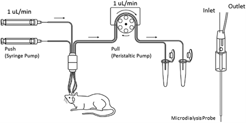
Figure 5. The Push-Pull system for large-pore microdialysis. The system requires both a syringe pump and a peristaltic pump to maintain a consistent flow rate before and after the microdialysis probe. The microdialysis probe has an open vent to keep the inner fluid pressure constant and prevents ultrafiltration of perfusion fluid from the probe into the brain tissue. More details have been discussed in Ref 2
Supplemental material

Supplemental Material

Download MS Word (369.8 KB)