Figures & data
Figure 1 (See previous page). Nwk-induced cellular protrusions do not depend on actin or microtubules for stability once formed. (A) Time-lapse imaging of S2 cells co-transfected with Lifeact-mCherry and either EGFP (left) or Nwk1-428-EGFP (right), and treated with Lat B after spreading on ConA. F-actin disassembles upon treatment, but the cellular protrusions remain. Images show 2D projections of confocal stacks. Scale bar, 10 µm. Time scale is in minutes. (B) Confocal microscopy of fixed S2 cells expressing EGFP or Nwk1-428-EGFP, and mCherry-tubulin. Nwk and tubulin localize to protrusions. Images show 2D projections of confocal stacks. Scale bar, 10 µm. Scale bar in magnified image is 5 µm. (C) Electron microscopy of Nwk1-428-EGFP–expressing S2 cells treated with Lat B to disrupt the actin cytoskeleton. Microtubules can be observed throughout the protrusion (arrows). Scale bar, 100 nm. (D) Live cell imaging of S2 cells expressing EB1-GFP with mCherry (top) or Nwk1-428-mCherry (bottom). EB1, a plus end microtubule binding protein, can be seen throughout protrusions (arrows, see also Movies 1 and 2). Scale bar, 10 µm. (E) Nwk-induced protrusions remain stable after microtubule depolymerization. Live cell imaging of S2 cells expressing Nwk1-428-mCherry and treated with Nocodazole, a microtubule-depolymerizing drug, or DMSO control. Images show 2D projections of confocal stacks taken before (t = 0), and 15 minutes after (t = 15) treatment. (F) Quantification of cell shape before and after treatment. Protrusion index is represented by cell perimeter divided by the square root of cell area (P/√A).
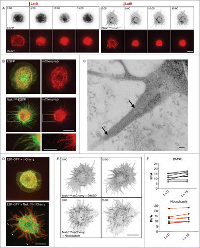
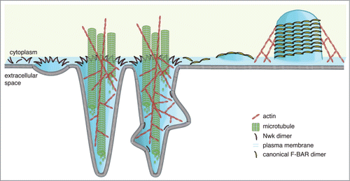
Figure 2. Model for formation of Nwk-induced protrusions at S2 cell plasma membranes. Higher-order assembly of Nwk in zigzags on the lipid bilayer results in membrane ridging and resultant inter-ridge buds or scallops (left), which are amplified by the cytoskeleton to form large-scale actin- and microtubule-filled cellular projections (center). In contrast, canonical F-BAR domains oligomerize into filaments and helical scaffolds to induce intracellular tubules (right).