Figures & data
Figure 1. In vitro analyses of vps1 E461K. (A) Structure of Dynamin-like protein- (pdb protein 4BEJ) with the residue equivalent to E461K (E426) highlighted on one of the helices of the dynamin stalk domain. (B) Actin binding of Vps1 wild type and the E461K mutant was assessed by incubating with F-actin followed by high speed centrifugation. The proportion of Vps1 found in the pellet in the presence and absence of actin is shown. Error bars are standard deviation. The proportion of actin in the pellet at high speed is not significantly different. (C) Wild type and mutant Vps1 were purified and analyzed using transmission electron microscopy following negative staining. For wild type, images shown are single and double rings in the absence of actin, and the double rings that form in the presence of actin filaments. For the E461K mutant images of 4 single rings are shown in the absence of actin and then rings in the presence of actin. No double rings were observed.
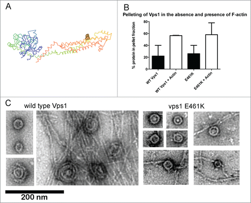
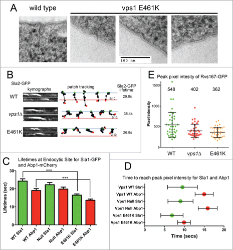
Figure 2. Analysis of cells expressing vps1 E461K. (A) Cells expressing wild type or vps1 E461K were high pressure frozen as previously described. Thin sections were imaged to observe plasma membrane invaginations. Examples of representative invaginations are shown. (B) Cells expressing wild type or vps1 E461K in the presence of the endocytic reporter Sla2-GFP were analyzed in live cells. Movies were recorded of endocytic patch movement and used to generate kymographs shown in the left panel. Patches were tracked and the ability of the patch to track further than 200 nm was recorded. The blue line denotes the plasma membrane while the red line is 200 nm into the cell from this point. The green spot indicates starting point, and the red spot the point of disassembly or where the patch moved away from the focal plane. Ten patches were analyzed for each and the proportion crossing the line noted. The lifetime of ≥30 patches for each strain was analyzed and noted on the right. (C) Endocytic patches detected with endocytic reporters Sla1-GFP and Abp1-mCherry were analyzed in wild type or vps1 E461K cells. Movies of cells were recorded and used to measure lifetimes of the reporters in 30 patches. Shown is lifetime ±SEM. P value <0 .0001 (***) from Students t tests indicates lifetimes for both Sla1 and Abp1 are significantly shorter that corresponding wild type values. (D) To determine whether E461K affects recruitment times at the endocytic site Sla1-GFP and Abp1-mCherry were analyzed in co-expression studies in live cells. At 30 individual sites the time taken for each marker to reach peak intensity was recorded. The green or red spot marks the mean time, and the error bar denotes range. (E) The peak pixel intensity of ≥37 patches of Rvs167-GFP were analyzed in wild type, vps1 null or vps1 E461K expressing cells. Numbers indicated are the mean intensity. E461K intensity is lower than that in wild type cells (p = 0.002 in students' t-test).