1,027
Views
8
CrossRef citations to date
0
Altmetric
Short Communication

Cyanobacterial Mn-catalase ‘KatB’: Molecular link between salinity and oxidative stress resistance

, , &
Article: e1216738 | Received 06 Jun 2016, Accepted 19 Jul 2016, Published online: 17 Aug 2016

Figures & data

Figure 1. Detection of oxidized proteins. Proteins were extracted by TCA precipitation from the culture medium of NaCl-treated wild-type Anabaena (WT) or the katB mutant (katB) cells after exposure to H2O2 (1mM). These proteins were derivatized with dinitrophenol (DNP), resolved on SDS-PAGE and transferred to nitrocellulose membrane. Subsequently, these proteins were probed with the monoclonal DNP antiserum. A Ponceau S-stained part of the blot is shown in the lower panel as loading control. The oxidized proteins were detected as mentioned in the OxyBlot oxidized protein detection kit (Thermo Scientific, 23280).

Figure 1. Detection of oxidized proteins. Proteins were extracted by TCA precipitation from the culture medium of NaCl-treated wild-type Anabaena (WT) or the katB mutant (katB−) cells after exposure to H2O2 (1mM). These proteins were derivatized with dinitrophenol (DNP), resolved on SDS-PAGE and transferred to nitrocellulose membrane. Subsequently, these proteins were probed with the monoclonal DNP antiserum. A Ponceau S-stained part of the blot is shown in the lower panel as loading control. The oxidized proteins were detected as mentioned in the OxyBlot oxidized protein detection kit (Thermo Scientific, 23280).

Figure 2. Ultrastructural features of the NaCl-treated wild-type Anabaena (WT, upper panel) or katB mutant (katB, lower panel) after exposure to H2O2 (for 24 h) as seen under the transmission electron microscope. Samples were processed for transmission electron microscopy as described earlier.Citation22 Thylakoid membranes (Th) and carboxysomes (C) are indicated. Severely disintegrated thylakoid membranes and a distinct loss of ultrastructure are evident in the katB mutant filaments exposed to H2O2.
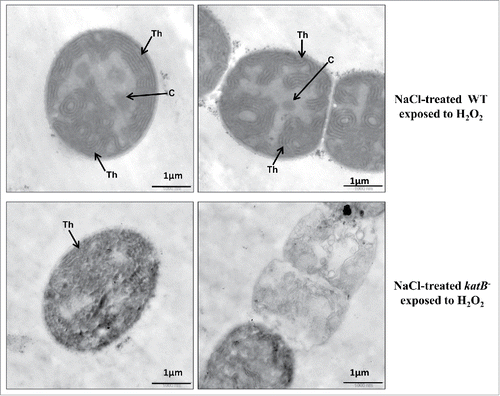
Figure 2. Ultrastructural features of the NaCl-treated wild-type Anabaena (WT, upper panel) or katB mutant (katB−, lower panel) after exposure to H2O2 (for 24 h) as seen under the transmission electron microscope. Samples were processed for transmission electron microscopy as described earlier.Citation22 Thylakoid membranes (Th) and carboxysomes (C) are indicated. Severely disintegrated thylakoid membranes and a distinct loss of ultrastructure are evident in the katB mutant filaments exposed to H2O2.

Figure 3. The katB promoter-gfp fusion construct was transformed into Anabaena PCC 7120 and the katB promoter activity was monitoredCitation13 in the presence of various osmolytes such as NaCl (150 mM), sucrose (300 mM), glycerol (300 mM), manitol (300 mM), sorbitol (300 mM), and PEG (100 mM). Cells were exposed to the above-mentioned osmolytes for 18h. The green fluorescence (λex = 490 nm, λem = 520 nm) of the reporter GFP is plotted as bar diagram. Standard deviation for 5 independent experiments is shown as error bars.
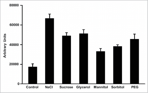
Figure 3. The katB promoter-gfp fusion construct was transformed into Anabaena PCC 7120 and the katB promoter activity was monitoredCitation13 in the presence of various osmolytes such as NaCl (150 mM), sucrose (300 mM), glycerol (300 mM), manitol (300 mM), sorbitol (300 mM), and PEG (100 mM). Cells were exposed to the above-mentioned osmolytes for 18h. The green fluorescence (λex = 490 nm, λem = 520 nm) of the reporter GFP is plotted as bar diagram. Standard deviation for 5 independent experiments is shown as error bars.