
Figures & data
Figure 1. (a) Comparison of a classic textbook representation of a nerve spike and (b) most probable structure in reality as per state-of-the-art studies.
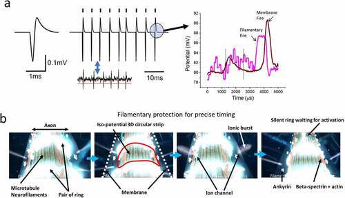
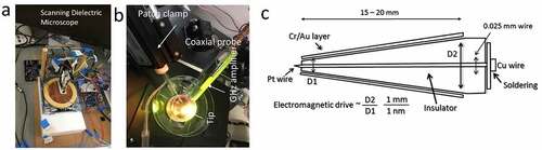
Figure 2. (a) Dielectric resonance microscope: biomaterials are kept on a metal or conducting plate. The tip scans through the surface, sending electromagnetic signals of various frequencies to the material and receiving the returned signal post-metal sheet reflection. (b) Coaxial probe and patch clamps are used together while measuring the filamentary firing and membrane firing. (c) The design of a coaxial probe.
Supplemental material