Figures & data
Figure 1. Rhizoid growth in the vertical culture method. (a) Vertical culture method. Gemmalings are grown vertically in a culture dish filled with ½ B5 medium (1% agar). Rhizoids grown over the surface of medium are examined. (b) Four-day old gemmalings of the wild type and Mpnek1 mutant. Photographed with a stereomicroscope S8APO0 (Leica Microsystems) equipped with a CCD camera DFC500 (Leica). (c) Sixteen-day old wild type and Mpnek1 mutant, photographed with a digital single-lens reflex camera (Nikon D5600). (d) Quantification of the rhizoid lengths of the wild type and Mpnek1 mutant (Male wild type: Tak-1, Female wild type: Tak-2, Male Mpnek1 mutant: line #2-4, Female Mpnek1 mutant: line #43). Values are shown by box plots (n = 26–47). Asterisks indicate significant difference compared with the wild type (Student's t-test, *P < 0.05).
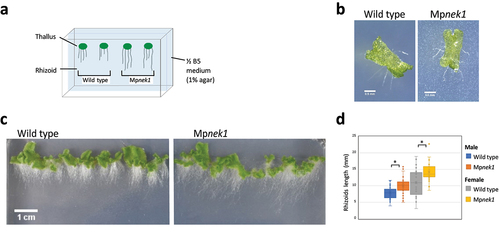
Figure 2. Rhizoid growth in the cellophane culture method. (a) Cellophane culture method. Gemmalings are grown vertically on a cellophane sheet placed on the surface of a ½ B5 medium (1% agar). The cellophane sheet hinders rhizoids from getting into the medium. Rhizoids grown on the surface of medium are examined. (b) Four-day old gemmalings of the wild type and Mpnek1 mutant. Photographed with a stereomicroscope S8APO0 (Leica Microsystems) equipped with a CCD camera DFC500 (Leica). (c) Sixteen-day old wild type and Mpnek1 mutant, photographed with a digital single-lens reflex camera (Nikon D5600). (d) Quantification of the rhizoid lengths in the wild type and Mpnek1 mutant (Male wild type: Tak-1, Female wild type: Tak-2, Male Mpnek1 mutant: line #2-4, Female Mpnek1 mutant: line #43). Values are shown by box plots (n = 40). No significant difference is observed (Student's t-test, P > 0.05).

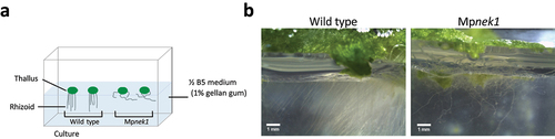
Figure 3. Rhizoid growth in the gellan-gum culture method. (a) Gellan-gum culture method. Gemmalings are grown on the ½ B5 medium solidified in the bottom half of a square culture dish by 1% gellan gum, which is more transparent than 1% agar and is suitable for lateral observation. Rhizoids grown inside the medium are examined. (b) Rhizoids in the 29-day-old of wild type and Mpnek1 mutant. Photographed with a stereomicroscope S8APO0 (Leica Microsystems) equipped with a CCD camera DFC500 (Leica).