
Figures & data
Table 1. The 2D and 3D structures of ligands.
Figure 1. Position of the redocked ligand (Purple) with the crystallographic ligand (Green) of the donepezil crystallographic ligand.
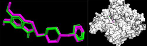
Table 2. Binding free energy and amino acid residues of ligands.
Figure 2. Donepezil and dieckol mechanism of action.
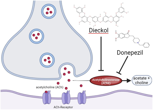
Figure 3. Fucodiphlorethol G (a); 7-Phloroecol (b); Dieckol (c); Fucoidan (d); Eckol (e); Laminaran (f); Alginic Acid (g); Donepezil (h).
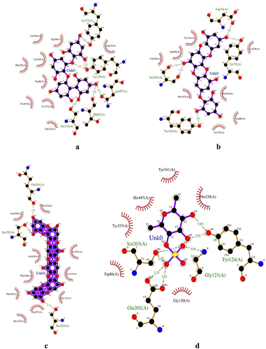
Figure 3. (Continued).
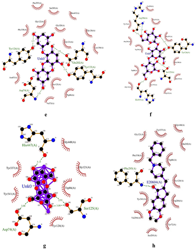
Table 3. Safety and toxicity results of Dieckol.
Data availability statement
The data generated during the study is available at https://doi.org/10.6084/m9.figshare.25765011.