

Figures & data
Figure 1. Root canal configurations by micro ct observed in mandibular premolars.
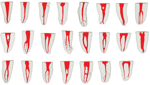
Figure 2. Vertucci’s classification by micro ct observed in mandibular premolars.
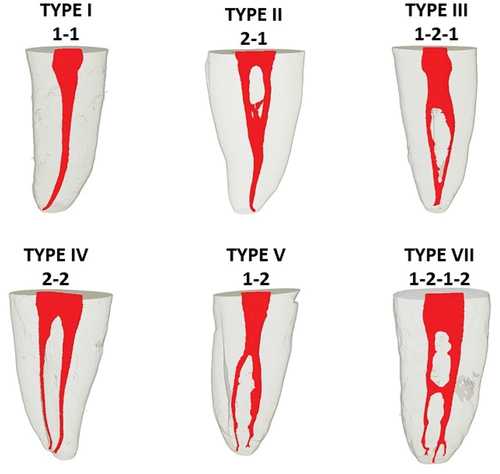
Table 1. Vertucci classification and ramifications of the root canal morphology of mandibular premolars evaluated by micro-computed tomography.
Figure 3. Anatomical variations and ramifications of mandibular premolars using micro-ct. (a).Lateral canal. (b). Secondary canal. (c). Apical delta. (d). Intercanal.
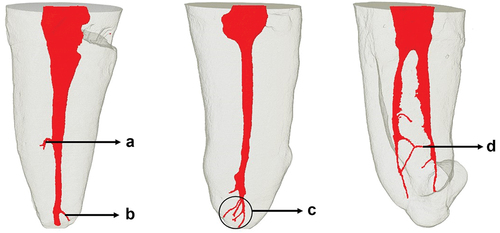
Table 2. Data from parameters evaluated at total and 5 mm from the apical foramen of mandibular premolars evaluated by micro-computed tomography.