

Figures & data
Table 1. Clinicopathological profile of the 40 parotid cases.
Table 2. Histologic profile of the 40 parotid cases and their grading according to the AFIP system.
Figure 1. Micrograph showing (a) Immunoreactive deposits appeared as reddish-brown reactions where the target antigen was located (MENA) in conventional low-grade MEC (40×); (b) Negative immunostaining for CD133 in low-grade oncocytic MEC (40×); (c) Negative immunostaining for CD44 in low-grade oncocytic MEC (40×). (d) Negative immunostaining for OCT4 in high-grade solid MEC (10×).
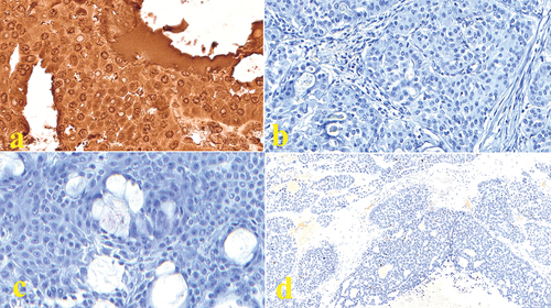
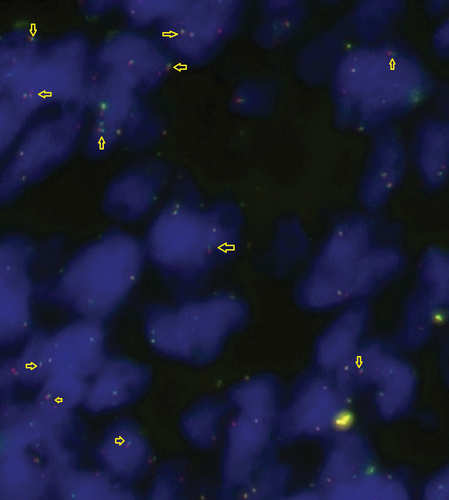
Figure 2. Photomicrograph showing retained signals for POU5F1 FISH probe.
Supplemental material