Figures & data
FIGURE 1 Relative head kidney lymphocyte and granulocyte counts in common carp following PCP exposure for 24 and 72 h. Values are given as mean ± SEM. P values provided are the probability that there are no differences between control and treatment groups. *P <.05.
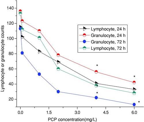
FIGURE 2 Mean EROD activity of common carp exposed to PCP for 24 and 72 h. Error shown is stranded error of the mean. A, control; B–F, 0.1, 1.0, 2.0, 4.0, and 6.0 mg/L PCP, respectively. *Significantly different from control value (P <.05).
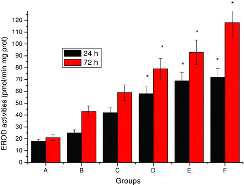
FIGURE 3 Mean GSH content of common carp exposed to PCP for 24 and 72 h. Error shown is stranded error of the mean. A, control; B–F, 0.1, 1.0, 2.0, 4.0, and 6.0 mg/L PCP, respectively. *Significantly different from control value (P <.05).
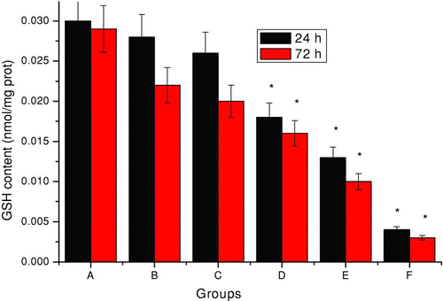
FIGURE 4 Mean MDA content of common carp exposed to PCP for 24 and 72 h. Error shown is stranded error of the mean. A, control; B–F, 0.1, 1.0, 2.0, 4.0, and 6.0 mg/L PCP, respectively. *Significantly different from control value (P <.05).
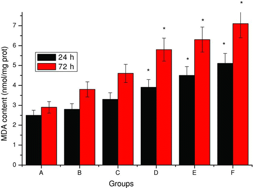
FIGURE 5 Effects of PCP on the mitochondrial transmembrane potential. Data are presented as means ± SD (n = 6). A, control; B–F, 0.1, 1.0, 2.0, 4.0, and 6.0 mg/L PCP, respectively. *Significantly different from control value (P <.05).
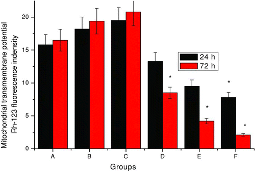
FIGURE 6 Mean concentration of testosterone in male common carp exposed to PCP for 24 and 72 h. Error shown is stranded error of the mean. A, control; B–F, 0.1, 1.0, 2.0, 4.0, and 6.0 mg/L PCP, respectively. ast;Significantly different from control value (P <.05).
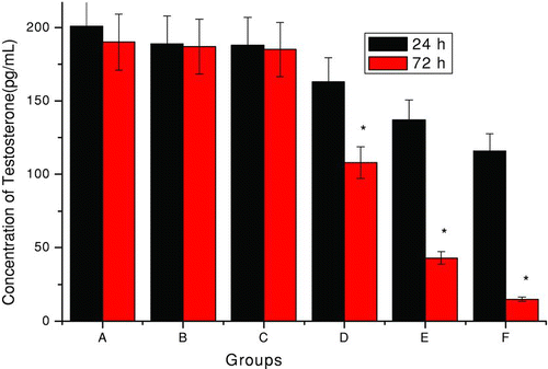
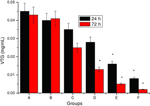
FIGURE 7 Plasma VTG in female common carp with 24 and 72 h of PCP exposure. Error shown is stranded error of the mean. A, control; B–F, 0.1, 1.0, 2.0, 4.0, and 6.0 mg/L PCP, respectively. *Significantly different from control value (P <.05).