Figures & data
Figure 1. Structure of leucomalachite green (LMG), malachite green (MG) and structurally similar compounds, leucocrystal violet (LCV), crystal violet (CV) and paraosaniline, used to determine cross-reactivity.
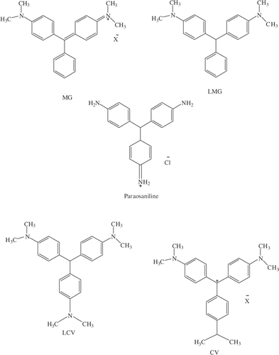
Figure 2. Synthetic scheme for the synthesis of LMG hapten and subsequent formation of immunogen by conjugation to a carrier protein (BSA).
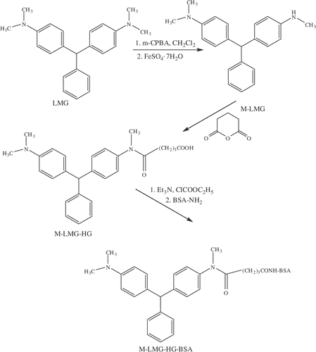
Figure 3. Synthetic scheme for the conjugation of LMG hapten to HRP.
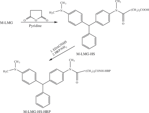
Figure 4. Representative calibration curve for LMG prepared in blank tilapia extract.
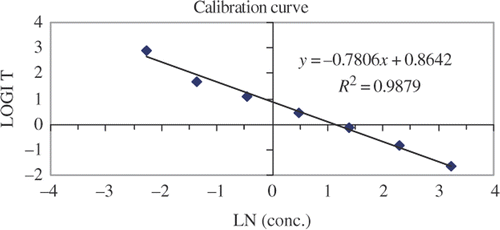
Table 1. Calibration curve results over a 3-day period with tilapia extract IC50, IC80 and IC90 in ng (LMG) g−1 tissue.
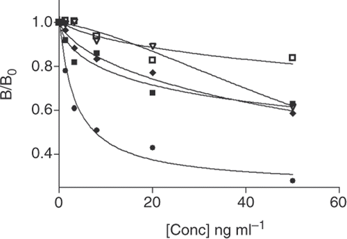
Figure 5. Calibration curves used to determine the cross-reactivity for LMG (•), MG (▪), LCV (♦), CV (□) and PS (▽).