9,386
Views
21
CrossRef citations to date
0
Altmetric
Articles

Biodesalination: an emerging technology for targeted removal of Na+ and Cl from seawater by cyanobacteria

, , , , , , , , , & show all
Pages 2647-2668 | Received 14 Apr 2014, Accepted 16 Jun 2014, Published online: 13 Oct 2014

Figures & data

Fig. 1. Literature searches indicating the specific growth rate of different cyanobacteria, algae and mixed environmental samples. In the figure above, it is evident that cyanobacteria appeared to have overall higher growth rates, when compared to algae.

Fig. 1. Literature searches indicating the specific growth rate of different cyanobacteria, algae and mixed environmental samples. In the figure above, it is evident that cyanobacteria appeared to have overall higher growth rates, when compared to algae.

Fig. 2. Physiological adaptations of cyanobacteria to ionic stress caused by Na+. The mechanisms described above give a brief overview of early responses, as well as long-term adaptations to high salinity.

Fig. 2. Physiological adaptations of cyanobacteria to ionic stress caused by Na+. The mechanisms described above give a brief overview of early responses, as well as long-term adaptations to high salinity.

Fig. 3(a). Effect of intermittent light in the growth of selected cyanobacteria. Data indicated that Synechococcus PCC 7002 was growing faster than Synechocystis PCC 6803 in all light intensities tested. In contrast, Synechocystis PCC 6803 appeared to show better overall growth under high light intensities, when compared to Synechococcus PCC 7002. Error bars indicate standard deviation (n = 3).

Fig. 3(a). Effect of intermittent light in the growth of selected cyanobacteria. Data indicated that Synechococcus PCC 7002 was growing faster than Synechocystis PCC 6803 in all light intensities tested. In contrast, Synechocystis PCC 6803 appeared to show better overall growth under high light intensities, when compared to Synechococcus PCC 7002. Error bars indicate standard deviation (n = 3).

Fig. 3(b). Effect of continuous light in the growth of selected cyanobacteria. Both Synechococcus PCC 7002 and Synechocystis PCC 6803 appeared to reach maximum growth on day 21 (week 3). Moreover, Synechococcus PCC 7002 grew best at lower light intensities, while Synechocystis PCC 6803 showed faster growth under high light intensities. It is worth noting that the maximum cell densities achieved was ten-fold lower than when grown under intermittent light. Error bars indicate standard deviation (n = 3).

Fig. 3(b). Effect of continuous light in the growth of selected cyanobacteria. Both Synechococcus PCC 7002 and Synechocystis PCC 6803 appeared to reach maximum growth on day 21 (week 3). Moreover, Synechococcus PCC 7002 grew best at lower light intensities, while Synechocystis PCC 6803 showed faster growth under high light intensities. It is worth noting that the maximum cell densities achieved was ten-fold lower than when grown under intermittent light. Error bars indicate standard deviation (n = 3).

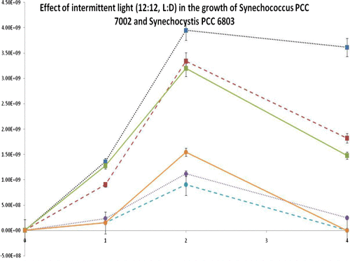
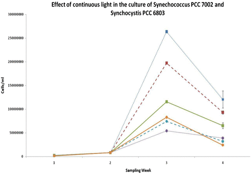
Table 1 Divisions per day as calculated by flow cytometry. The table below demonstrates growth rate (per day) for Synechococcus PCC 7002 and Synechocystis PCC 6803, under different illuminations. Red indicates highest growth observed for each organism over 3 weeks of culture. Green indicates lowest growth for each organism per period.

Fig. 4. HPLC detection standard curve for Na+ and Cl. The limits of quantification for both equations were limited between 10 and 100 mM. Error bars indicate standard deviation (n = 3).

Fig. 4. HPLC detection standard curve for Na+ and Cl−. The limits of quantification for both equations were limited between 10 and 100 mM. Error bars indicate standard deviation (n = 3).

Fig. 5. XPS analysis of Synechocystis PCC 6803 and Synechococcus PCC 7002. Values denote the distribution of macromolecules on the cell surface in the analysed sample volume. Error bars are standard deviation values.

Fig. 5. XPS analysis of Synechocystis PCC 6803 and Synechococcus PCC 7002. Values denote the distribution of macromolecules on the cell surface in the analysed sample volume. Error bars are standard deviation values.

Fig. 6. XDLVO analysis. Aggregation energies as a function of separation distance for (A) Synechocystis PCC 6803 and (B) Synechococcus PCC 7002. G—interaction energy, GLW—interaction energy due to Lifschitz–van der Waals component, GEL—interaction energy due to electrostatic component, GAB—interaction energy due to acid–base component, GTOT—total interaction energy. Insets show the formation of the predicted secondary minimum at which reversible cell aggregation might occur.

Fig. 6. XDLVO analysis. Aggregation energies as a function of separation distance for (A) Synechocystis PCC 6803 and (B) Synechococcus PCC 7002. G—interaction energy, GLW—interaction energy due to Lifschitz–van der Waals component, GEL—interaction energy due to electrostatic component, GAB—interaction energy due to acid–base component, GTOT—total interaction energy. Insets show the formation of the predicted secondary minimum at which reversible cell aggregation might occur.