Figures & data
Top view and higher magnification (inset) images from a scanning electron microscope of an ordered hollow urchin-like structure of ZnO nanowires [Citation11]. Courtesy of Dr. J. Elias (EMPA Materials Science and Technology).
![Top view and higher magnification (inset) images from a scanning electron microscope of an ordered hollow urchin-like structure of ZnO nanowires [Citation11]. Courtesy of Dr. J. Elias (EMPA Materials Science and Technology).](/cms/asset/356a0ebf-b8a7-4e91-bf18-b21565c594b3/tsnm_a_663812_o_f0001g.gif)
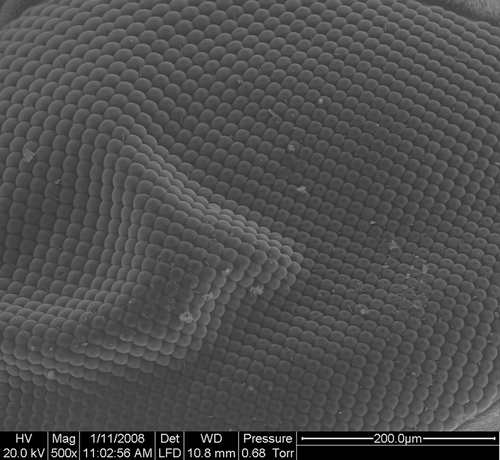
Scanning electron microscope image of the compound eye of a fly.
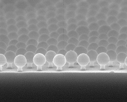
Tilted image on a scanning electron microscope of a templating array of 360-nm-diameter spheres of silica and the silicon nipples etched underneath. Courtesy of Prof. P. Jiang (University of Florida).
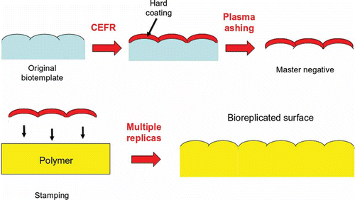
Schematic of the Nano4Bio technique.