Figures & data
Figure 1. (a) TEM image [Citation1] of Co80Ni20 nanoparticles with 50 nm average diameter and (b) SEM image [Citation13] of the Co80Ni20 nanoparticles prepared by polyol process.
![Figure 1. (a) TEM image [Citation1] of Co80Ni20 nanoparticles with 50 nm average diameter and (b) SEM image [Citation13] of the Co80Ni20 nanoparticles prepared by polyol process.](/cms/asset/9c611259-18fc-4a87-85ae-7b13c2b0c0f0/tsnm_a_672343_o_f0001g.gif)
Figure 2. (a) Hysteresis loops of the Co80Ni20 nanocomposite at selected temperatures from 5 K to 300 K after correction for the diamagnetic PVC matrix. Main panel: highlight of the hysteresis loops over field range –0.5 to 0.5 Tesla. Inset: hysteresis loops over the field range –5 to 5 Tesla. (b) Highlight of hysteresis loops in (a) over the field range –0.2 to 0.0 Tesla. The arrow illustrates the shift of decreasing coercivity H c (increasing –H c) as the temperature increases from 5 K to 300 K.
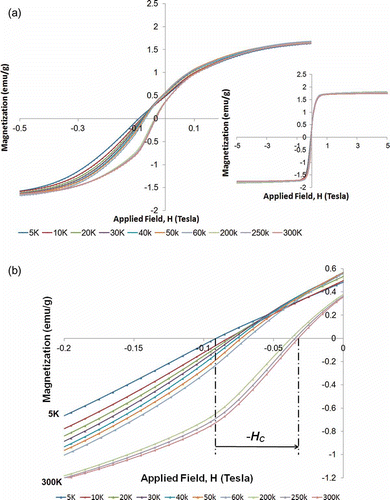
Figure 3. Applied field dependence of irrversible susceptibility for the Co80Ni20 nanocomposite at selected temperatures from 5 K to 300 K. The maximum irreversible susceptibility χ irr,max of each curve (peak value) is represented by black dot. The arrow illustrates the shift of increasing χ irr,max as the temperature increases from 5 K to 300 K.
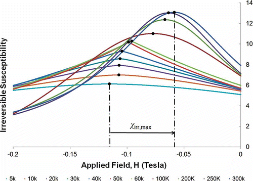
Figure 4. Applied field and temperature dependence of irreversible susceptibility for the Co80Ni20 nanocomposite. The color varies from light green for small values of χ irr to purple for χ irr,max.
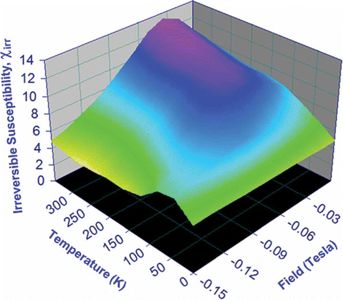
Figure 5. Extraction of decay coefficient from decay curve using MPA model.
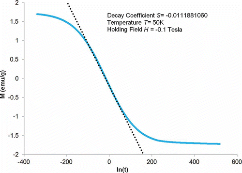
Figure 6. Holding field dependence of decay coefficient, S, for the Co80Ni20 nanocomposite at selected temperatures from 10 K to 300 K. As temperature increases from 10 K to 300 K, S max increases first from 10 K to 50 K, reaches peak value at 50 K and then decreases from 50 K to 300 K.
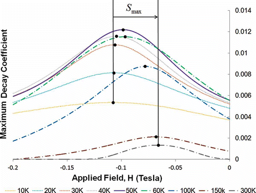
Figure 7. Temperature dependence of the maximum decay coefficient for the Co80Ni20 nanocomposite. MPA model evaluated S max at selected temperatures are represented by red dots. The non-Arrhenius behavior is illustrated by the black fitting curve.
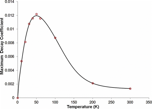
Figure 8. Temperature and field dependence of the maximum decay coefficient for the Co80Ni20 nanocomposite. The maximum decay coefficient varies from light green for small values of S max to dark blue for peak value of S max.
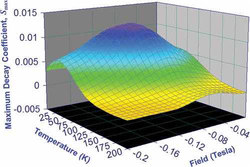
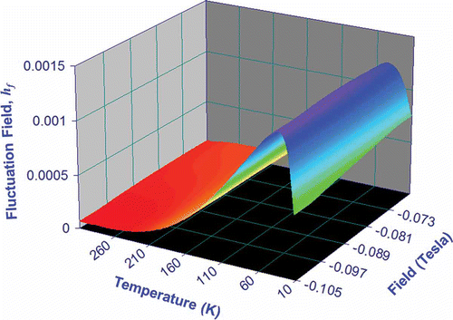
Figure 9. Temperature and applied field dependence of the fluctuation field for the Co80Ni20 nanocomposite. hf were rigorously determined from macroscopic measurements of S and χ irr .
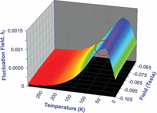
Figure 10. Temperature dependence of fluctuation field at a representative applied field of –0.1 Tesla.
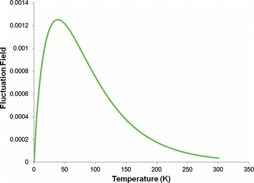
Figure 11. Temperature and applied field dependence of the fluctuation field for the Co80Ni20 nanocomposite. hf
were evaluted using EquationEquation (7)(7).