Figures & data
Figure 1. AFM images of non-dialyzed SDS-SWNT hybrids on AP-mica. (a) An un-rinsed sample. (b) A rinsed sample. Scan size of the images was 2 μm × 2 μm.
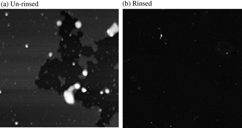
Figure 2. Schematic views of experiment. (a) Dialysis procedure. (b) Sample preparation for AFM observations.
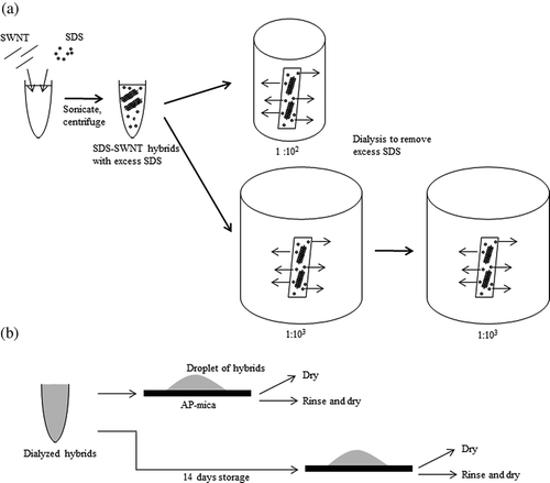
Figure 3. AFM images of dialyzed SDS-SWNT hybrids on AP-mica. (a, d) The volume ratio of dispersion to water in dialysis was 1:102. (b, e) The volume ratio of dispersion to water in dialysis was 1:103. (c, f) The volume ratio of dispersion to water in dialysis was 1:106. Cases (a) to (c) and (d) to (f) were not rinsed and rinsed with water before drying, respectively. Lines in figures indicate cross sections of SDS-SWNT hybrids. Scan size of the images was 2 μm × 2 μm.
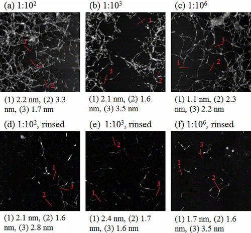
Figure 4. Magnified AFM images of dialyzed SDS-SWNT hybrids on AP-mica. (a, d) The volume ratio of dispersion to water in dialysis was 1:102. (b, e) The volume ratio of dispersion to water in dialysis was 1:103. (c, f) The volume ratio of dispersion to water in dialysis was 1:106. Cases (a) to (c) and (d) to (f) were not rinsed and rinsed with water before drying, respectively. Scan size of the images was 500 nm × 500 nm.
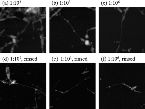
Figure 5. AFM images of SDS-SWNT hybrids stored for 14 days after dialysis. (a), (b), and (c) The volume ratios of dispersion to water in dialysis were 1:102, 1:103, and 1:106, respectively. Scan size of the images was 2 μm × 2 μm.
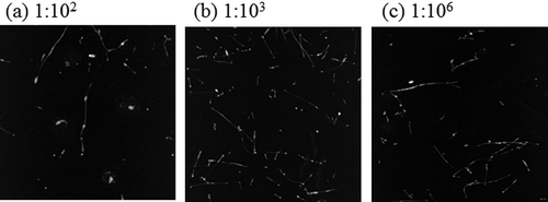
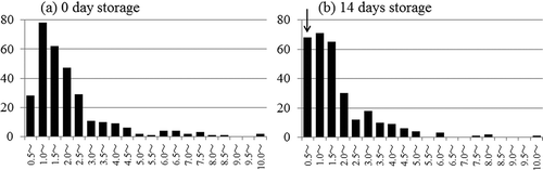
Figure 6. Histograms of diameter distribution of SDS-SWNT hybrids. (a) Dialyzed hybrids were observed immediately after dialysis. (b) Dialyzed hybrids were stored for 14 days before AFM observations.