Figures & data
Figure 1. Disease induction of mice with oral injecting Bacillus megaterium expressing toxins. Mice were inoculated with live Bacillus megaterium bacterial cells expressing wild-type TcdB (108 bacteria/injection), aTcdB (109), or GFP (109) twice a day for 10 d. Mouse weights (A) and overall survival (B) were monitored over time. The statistical analysis of TcdB in comparison with GFP group. * represents p < 0.05 whereas ** shows p < 0.01 and *** shows p < 0.001. aTcdB group showed no statistical significance in either weight loss or survival curves when compared with GFP group.
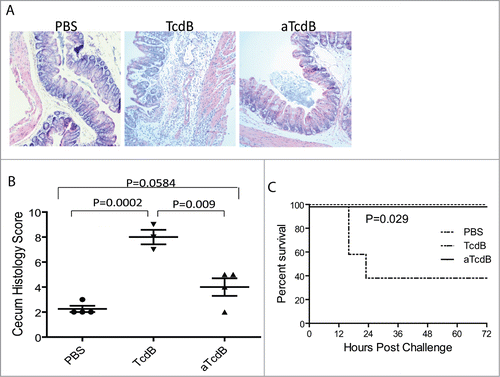
Figure 2. Disease induction of mice with cecum injection of wild type and mutant toxins. (A) Histology of cecum tissues from mice injected with PBS, TcdB, or aTcdB. (B) Histology scores of cecum tissue sections. P values between the groups were shown. (C) Survival curves of mice after toxin cecum injection. P < 0.05 between TcdB and PBS groups.