Figures & data
Table 1. ELISA results to assess H. pylori status in mothers using cord blood from their children or breastmilk.
Table 2. Serologic markers of H. pylori status in 105 birth-cohort children at 24 months of age.
Table 3. Relationship between maternal H. pylori status and child's H. pylori status at 24 months of age.
Table 4. Response to H. pylori antigens in 105 Bangladeshi birth-cohort children, at 24 months of age.
Table 5. Calculated seroconversion rates for H. pylori persistence in 105 Bangladeshi birth-cohort children during the first two years of life.
Figure 1. Western blot of H. pylori antigen preparations with human and rabbit serum samples. Panel A. Whole cell preparation of H. pylori 26695; Panel B. Water extracted whole cell antigen (WEWCA), as described.Citation20 Panel C. Recombinant CagA protein, as described.Citation83 Sera are: Lane 1, H. pylori+/CagA+ patient (H, human serum), lane 2, H. pylori+/CagA− patient. Lane 3, H. pylori−/CagA− patient, and Lane 4, serum from rabbit immunized with purified H. pylori CagA antigen (R, rabbit).
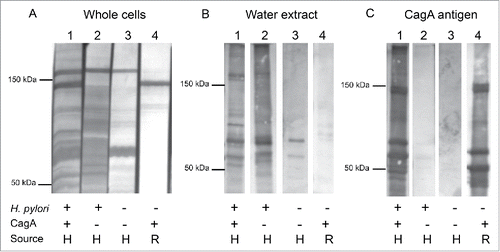
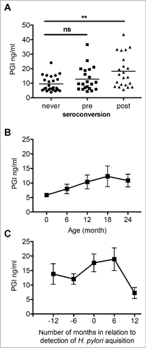
Figure 2. Serum pepsinogen I (PGI) levels of 18 birth-cohort children. Panel A. PGI levels of samples (n = 20) from children who never seroconverted compared to children with seroconversion (split into samples from children before (n = 21) and after (n = 23) seroconversion). Horizontal bars indicate the mean.**p = 0.001; Panel B. Serum PGI levels over time (mean±SEM ng/ml) in five children who were persistently H. pylori sero-negative and were followed from birth; n = 5 per time point. Panel C. Changes in PGI levels (mean±SEM ng/ml) associated with H. pylori seroconversion in children who became persistently sero-positive. n = 9, 12, 13, 9, 3 for the indicated time points respectively;