Figures & data
Figure 1. Experimental design.
(A) Germ-free Il10-/- mice (n = 6) were compared with Il10-/- mice colonized with A. muciniphila at the age of 8 weeks (n = 6). Three weeks after start of the experiment the mice were euthanized. (B) Germ-free lL-10-/- mice were colonized with E. coli NC101 at the age of 9 weeks (n = 6) and compared with mice additionally colonized with A. muciniphila one week before E. coli NC101 colonization (n = 6). Two weeks after E. coli NC101 colonization the mice were euthanized. (C) Il10-/- mice colonized with the SIHUMI consortium (n = 6) were compared to Il10-/- mice, which were additionally colonized with A. muciniphila at the age of 8 weeks (n = 6). Three weeks after start of the experiment the mice were euthanized.
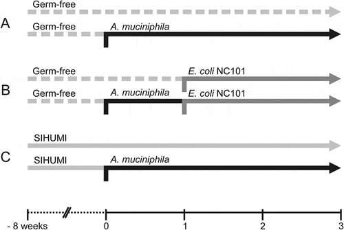
Figure 2. Mono-association of Il10-/- mice with A. muciniphila did not induce intestinal inflammation within three weeks.
(A) Body-weight development of A. muciniphila (Amuc) mono-associated Il10-/- mice within 3 weeks after colonization in comparison to germ-free (GF) Il10-/- mice. Mean ± SD, n = 6, *p < 0.05. (B) Cecal and colonic histopathology score of germ-free (GF) Il10-/- mice and Il10-/- mice mono-associated with A. muciniphila (Amuc). Horizontal dotted line: Maximal score. Median + range, n = 5–6, *p < 0.05. (C) Colon length of germ-free (GF) Il10-/- mice and Il10-/- mice mono-associated with A. muciniphila (Amuc). Mean ± SD, n = 5–6, *p < 0.05. (D) Relative mRNA levels of Tnfa, Ifng and Reg3g in mucosa of distal small intestine (SI), cecum and colon of Il10-/- mice mono-associated with A. muciniphila (Amuc) in comparison to germ-free (GF) Il10-/- mice. Mean ± SD, n = 3–4, *p < 0.05. (E) Fecal lipocalin-2 concentration of germ-free (GF) Il10-/- mice and Il10-/- mice mono-associated with A. muciniphila (Amuc). Median, n = 4–6, *p < 0.05, **p < 0.01.
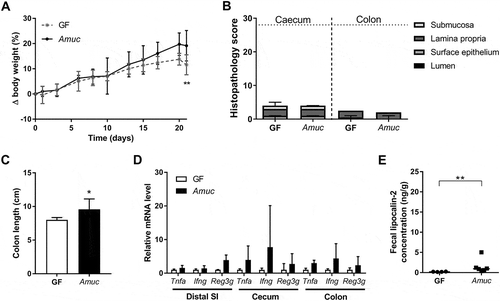
Figure 3. A. muciniphila does not modulate intestinal inflammation triggered by E. coli NC101 in Il10-/- mice.
(A) Body-weight development of originally germ-free and A. muciniphila (Amuc) mono-associated Il10-/- mice additionally colonized with E. coli NC101 (Ecoli) at day 7 (Vertical dotted line) in comparison to germ-free (GF) Il10-/- mice. Mean ± SD, n = 6, *p < 0.05, **p < 0.01, ***p < 0.001 compared to GF, #p < 0.05 for comparison between colonized Il10-/- mice. (B) Cecal and colonic histopathology score of germ-free (GF) Il10-/- mice, Il10-/- mice mono-associated with E. coli NC101 (Ecoli) and Il10-/- mice dual-associated with A. muciniphila and E. coli NC101 (Amuc + Ecoli). Horizontal dotted line: Maximal score. Median + range, n = 5–6, *p < 0.05. (C) Colon length of germ-free (GF) Il10-/- mice, Il10-/- mice mono-associated with E. coli NC101 (Ecoli) and Il10-/- mice dual-associated with A. muciniphila and E. coli NC101 (Amuc + Ecoli). Mean ± SD, n = 5–6, *p < 0.05. (D) Relative mRNA levels of Tnfa, Ifng and Reg3g in mucosa of distal small intestine (SI), cecum and colon of E. coli NC101 mono-associated Il10-/- mice and Il10-/- mice dual-associated with A. muciniphila and E. coli NC101 (Amuc + Ecoli) in comparison to germ-free (GF) Il10-/- mice. Mean ± SD, n = 4–6, *p < 0.05, **p < 0.01. (E) Fecal lipocalin-2 concentration of germ-free (GF) Il10-/- mice, Il10-/- mice mono-associated with E. coli NC101 (Ecoli) and Il10-/- mice dual-associated with A. muciniphila and E. coli NC101 (Amuc + Ecoli). Median, n = 4–6, *p < 0.05.
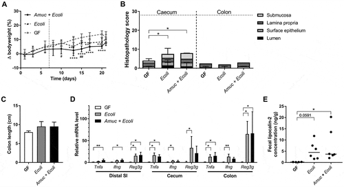
Figure 4. Colonization with A. muciniphila has no short-term effect on the colitogenic activity of the SIHUMI consortium in Il10-/- mice.
(A) Body-weight development of Il10-/- mice born with the SIHUMI consortium (SIHUMI) and SIHUMI-Il10-/- mice additionally colonized with A. muciniphila (SIHUMI + Amuc) in comparison to germ-free (GF) Il10-/- mice. Mean ± SD, n = 6, *p < 0.05 compared to GF, #p < 0.05, ##p < 0.01 for comparison between colonized Il10-/- mice. (B) Cecal and colonic histopathology score of germ-free (GF) Il10-/- mice, SIHUMI-Il10-/- mice (SIHUMI) and SIHUMI-Il10-/- mice additionally colonized with A. muciniphila (SIHUMI + Amuc). Horizontal dotted line: Maximal score. Median + range, n = 5–6, *p < 0.05, **p < 0.01. (C) Colon length of germ-free (GF) Il10-/- mice, SIHUMI-Il10-/- mice (SIHUMI) and SIHUMI-Il10-/- mice additionally colonized with A. muciniphila (SIHUMI + Amuc). Mean ± SD, n = 5–6, *p < 0.05. (D) Relative mRNA levels of Tnfa, Ifng and Reg3g in mucosa of distal small intestine (SI), cecum and colon of SIHUMI-Il10-/- mice (SIHUMI) and SIHUMI-Il10-/- mice additionally colonized with A. muciniphila (SIHUMI + Amuc) in comparison to germ-free (GF) Il10-/- mice. Mean ± SD, n = 4–6, *p < 0.05, **p < 0.01. (E) Fecal lipocalin-2 concentration of germ-free (GF) Il10-/- mice, SIHUMI-Il10-/- mice (SIHUMI) and SIHUMI-Il10-/- mice additionally colonized with A. muciniphila (SIHUMI + Amuc). Median, n = 4–6, *p < 0.05.
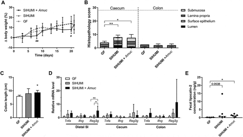
Figure 5. The cecal histology of gnotobiotic Il10-/- mice is not affected by A. muciniphila.
Histology of haematoxylin and eosin-stained cecal tissue sections from (A) germ-free (GF) Il10-/- mice, (B) Il10-/- mice mono-associated with A. muciniphila (Amuc) or (C) E. coli NC101 (Ecoli), (D) Il10-/- mice colonized with E. coli NC101 after pre-colonization with A. muciniphila (Amuc + Ecoli), (E) SIHUMI-Il10-/- mice (SIHUMI) and (F) SIHUMI-Il10-/- mice additionally colonized with A. muciniphila (SIHUMI + Amuc). Bar: 50 μm.
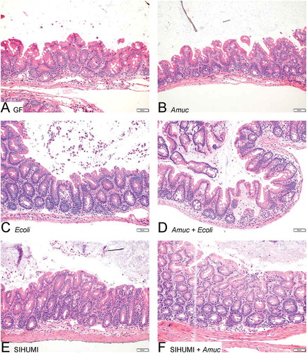
Table 1. Bacterial cell numbers in cecal contents of gnotobiotic Il10-/- mice.
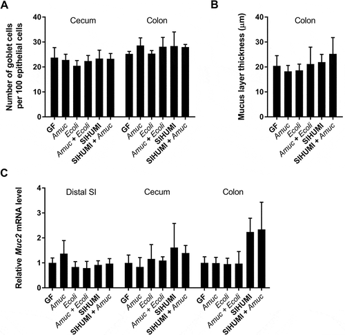
Figure 6. The mucus turnover in gnotobiotic Il10-/- mice is not influenced by A. muciniphila.
(A) Ratio of acidic mucin filled goblet cells in the cecal and colonic epithelium, (B) thickness of the colonic mucus layer and (C) relative mRNA levels of Muc2 in mucosa of distal small intestine (SI), cecum and colon of germ-free (GF) Il10-/- mice, Il10-/- mice mono-associated with A. muciniphila (Amuc) or E. coli NC101 (Ecoli), Il10-/- mice colonized with E. coli NC101 after pre-colonization with A. muciniphila (Amuc + Ecoli), SIHUMI-Il10-/- mice (SIHUMI) and SIHUMI-Il10-/- mice additionally colonized with A. muciniphila (SIHUMI + Amuc). Mean ± SD, n = 3–6, *p < 0.05.