
Figures & data
Figure 1. IL-27 induced LL-37 expression in primary human colonic epithelial cells. (a) Kinetic gene expression of LL-37 in primary human colonic epithelial cells after stimulation with different doses of recombinant human IL-27 protein (20, 50 and 100 μg/ml) for different times (6, 12 and 24 hours). (b) Representative Western blot analysis of LL-37 protein expression in cell lysates of primary human colonic epithelial cells at 24 hour after stimulation with different doses of recombinant human IL-27 protein (20, 50 and 100 μg/ml). Total proteins were extracted from primary human colonic epithelial cells (1 × 10⁶ cells), and an equal amount of protein (10 μg) was subjected to SDS-PAGE (10%) before blotting onto a PVDF membrane. β-Actin was used as a control to ensure an equal amount of loaded protein. (c) Effects of signaling molecule inhibitors on LL-37 protein expression. Primary human colonic epithelial cells were pretreated with AG490 (5 μM; AG), LY294002 (10 μM; LY), SB203580 (20 μM; LY), or U0126 (10 μM; U) for 1 hour, followed by incubation for 24 hour with or without IL-27 (50 ng/ml). LL-37 protein expression in cell lysates was analyzed by Western blot. Densitometry quantification of blots was shown as in histograms on the right. LL-37 expression was normalized to β-actin for each sample, and expression was graphed as fold change above cells at 0 min. Results are expressed as mean ± SD of 5 independent experiments. *P < .05, **P < .01,***P < .001 when compared between groups denoted by horizontal lines
Figure 2. Lack of IL-27 signaling impaired CRAMP expression in murine CDI. Wild-type (WT) and IL-27 receptor-deficient (WSX-1−/−) mice were infected with 104 colony-forming units of C. difficile VPI 10463 after antibiotic pretreatment. (a) At the indicated time after C. difficile infection, mice were sacrificed and ceca were harvested. Then total cellular RNA was extracted, and mRNA expression of CRAMP in colon tissues from mice (12 mice per group) was analyzed by quantitative PCR after C. difficile challenge. Details are described in the online supplementary methods. (b) Representative Western blot analysis of CRAMP protein expression in colon tissues from mice (12 mice per group) at day 0 or 1 after C. difficile challenge. (c) Immunohistochemistry by standard methods with anti-CRAMP antibody on fixed colon tissues of mice (12 mice per group) at day 0 or 3 after C. difficile challenge. Details are described in the online supplementary methods. Red arrows point to areas of CRAMP staining (brown). (d) CRAMP protein levels quantified by ELISA in the stools, colon tissues and sera of mice after C. difficile challenge. Details are described in the online supplementary methods. Results are expressed as mean ± SD of 3 independent experiments. *P < .05, ***P < .001 when compared between groups denoted by horizontal lines
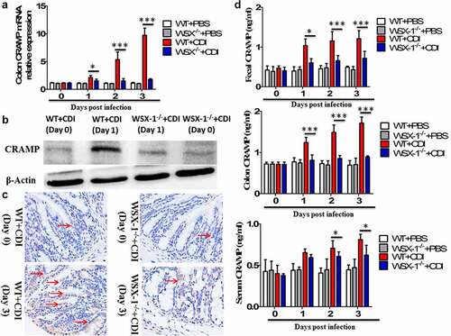
Figure 3. The effects of IL-27 administration or IL-27 blockade on CRAMP production. (a) CRAMP protein levels at day 1 in the colon tissues from mice (12 mice per group) treated with or without recombinant mouse IL-27 protein (0.5 μg/mice) immediately after C. difficile challenge. Phosphate buffer saline (PBS) was used as vehicle control. (b) CRAMP protein levels at day 1 in the colon tissues from mice (12 mice per group) treated with or without goat anti-IL-27 polyclonal IgG antibodies (100 μg/mice) immediately after C. difficile challenge. Polyclonal goat IgG was used as isotype control. Results are expressed as mean ± SD of 3 independent experiments. *P < .05, **P < .01 when compared between groups denoted by horizontal lines
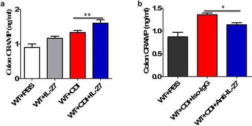
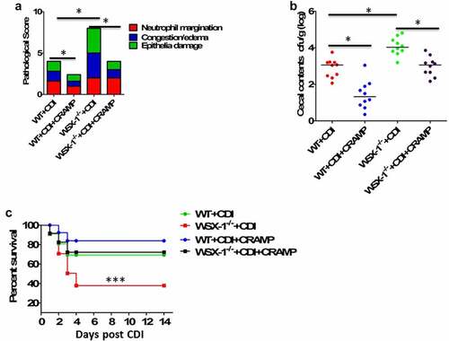
Figure 4. Exogenous CRAMP reduced C.difficile infection-associated mortality and morbidity in IL-27 receptor-deficient mice. WSX-1−/- mice were treated with or without CRAMP peptide 2 hours after infection with of C. difficile. (a) Tissue pathology scores of cecal tissues from WSX-1−/- mice (10 mice per group) treated with or without CRAMP peptide. Data were expressed as mean ± SD. *P < .05 when compared between groups denoted by horizontal lines. (b) C. difficile bacterial burden in cecal contents from WSX-1−/- mice (10 mice per group) on day 3 post C. difficile infection. Horizontal bars represent median values, and dots represent individual mouse. *P < .05 when compared between groups denoted by horizontal lines ((Mann–Whitney U test). (c) Survival was monitored until 2 weeks postinfection (12 mice per group). Comparison between groups was done by Kaplan-Meier analysis followed by log-rank tests. ***P < .001 when compared with WSX-1−/- mice treated with CRAMP peptide
Supplemental Material
Download Zip (2.1 MB)Availability of data
All data generated or analyzed during this study are included in this published article and its supplementary information files.