
Figures & data
Figure 1. Assays investigation of deacetylation of CobB and its role in virulence regulation.
(a) Western blot analysis of acetylation in protein lysates from WT, cobB mutant (ΔcobB) and cobB-overexpressing strains (cobB++). Coomassie brilliant blue was used as the loading control. (b) Adherence assays of WT and ΔcobB to HeLa cells. Data are presented as the mean ± SD (n = 3). (c) Detection of A/E lesion formation by WT and ΔcobB was evaluated by FAS in HeLa cells 3 hours post infection. The HeLa cell actin cytoskeleton (green) and nuclei of bacterial and HeLa cells (red) are shown. (d) FAS assay quantification of the number of pedestals per infected cell (n = 50). Data are presented as the mean ± SD (n = 3). (e) Competition assay comparing the colonization ability of ΔcobB and WT in the colon of rabbits. The competitive index (CI) is defined as the output ratio of ΔcobB to WT divided by the input ratio of WTkana to WTnadi. Each symbol represents the CI of an individual rabbit (n = 6). (f) Growth curves of WT and ΔcobB in DMEM. Data represent the mean ± SD (n = 3). Significant differences were assessed by an unpaired t test (b, d) or Mann – Whitney U test (e). Error bars represent SD. *p < .05, **p < .01, ***p < .001; n.s. no significant difference.
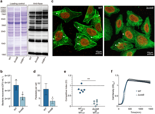
Figure 2. Identification of the substrates of CobB by using quantitative acetylome comparison of ΔcobB and WT.
(a) Venn diagram showing the overlap of acetylated sites compared between WT and ΔcobB. (b) A column chart showing all the acetylation sites increased or decreased in abundance in the WT and ΔcobB strains. The green column represents the sites increased in abundance. The blue column represents the sites decreased in abundance. FC = Fold change. (c) Gene Ontology analysis of acetylated proteins increased in abundance in ΔcobB, which are also protein candidates targeted by CobB. These proteins were analyzed for enrichment in three GO ontologies: biological process (upper), cellular component (middle), and molecular function (lower). The p value cutoff = 0.05 and q-value cutoff = 0.2 were selected as the cutoff criteria. Benjamini and Hochberg correction was used to adjust p values.
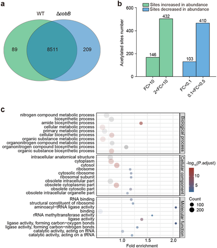
Figure 3. Assays of bacterial adhesion to HeLa cells and bacterial colonization of rabbits.
(a) Adhesion assays of the WT, ΔcesA, cesA+, cesA-K44Q, and cesA-K44R strains to HeLa cells. Data represent the mean ± SD (n = 3). (b) Detection of A/E lesion formation by WT, ΔcesA, cesA+, cesA-K44Q, and cesA-K44R by FAS of HeLa cells at 3 hours post infection. The HeLa cell actin cytoskeleton (green) and nuclei of bacterial and HeLa cells (red) are shown. (c) FAS assay quantification of the number of pedestals per infected cell (n = 50). (d) Colonization assays of the WT, ΔcesA, cesA+, cesA-K44Q, and cesA-K44R strains in the colon of rabbits (n = 6). Significant differences were assessed by an unpaired t test (a, c) or Mann – Whitney U test (d). Error bars represent SD. *p < .05, **p < .01, ***p < .001; n.s. no significant difference.
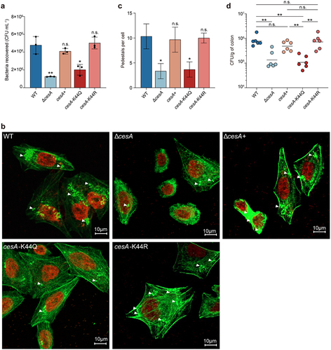
Figure 4. Assays of EspA stability and interaction with CesA.
(a) Western blot analysis of EspA stability in the cesA-K44Q and cesA-K44R backgrounds. (b) Western blot analysis of the EspA interaction with CesA-FLAG (WT), CesA-K44Q-FLAG (K44Q), and CesA-K44R-FLAG (K44R) purified by immunoprecipitation. The data are presented as the means ± SDs (n = 3). (c) Structural representation of the interactions between K44 of CesA and EspA. The left part is based on the X-ray crystal structure of the CesA-EspA complex (PDB: 1XOU). The right structure was generated through computational modeling. Significant differences were assessed by an unpaired t test. Error bars represent SD. *p < .05, **p < .01, ***p < .001; n.s. no significant difference.
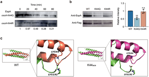
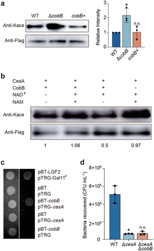
Figure 5. Assays of CesA interactions with CobB.
(a) Western blot and quantitative analysis of the acetylation level of the CesA-FLAG protein immunoprecipitated from strains cesA-Flag (WT), ΔcobBcesA-Flag (ΔcobB) and ΔcobB (+) cesA-Flag (ΔcobB+). “Anti-Kace” means “pan-anti-acetyllysine antibody”. (b) Western blot of CesA, incubated with CobB in the presence of NAD+ or NAM as inhibitor. Ten hours of incubation at 25°C revealed that NAD+-dependent CobB can catalyze deacetylation with CesA. (c) Bacterial two-hybrid assays for the interaction between CobB and CesA. Left panel: plate minus streptomycin (str) and 3-AT. Right panel: plate plus 12.5 mg/mL str and 15 mM 3-AT. Co-transformants containing pBT-LGF2 and pTRG-Gal11P were used as positive controls. A co-transformant containing pBT and pTRG was used as a negative control. Each unit representing the corresponding co-transformant in the plates is indicated in the figure. (d) Adhesion assays of the WT, ΔcesA, and ΔcesAΔcobB strains to HeLa cells. Data are presented as the mean ± SD (n = 3). Significant differences were assessed by an unpaired t test. Error bars represent SD. *p < .05, **p < .01, ***p < .001; n.s. no significant difference.
Supplemental material
0311_Revised_Supplementary Information.docx
Download MS Word (25.6 KB)0311_Revised_Supplementary Datasets.xlsx
Download MS Excel (1.7 MB)Data availability statement
The relevant data are provided in the manuscript (Supplementary Information). MS proteomics data were deposited in the ProteomeXchange Consortium (http://proteomecentral.proteomexchange.org) via the iProX partner repository with the dataset identifier PXD045950.