
Figures & data
Figure 1. A. Epithelium organization of the small intestine. Graphical representation of villi, crypts and different type of intestinal cells. B. Colon epithelium. Graphical representation of crypts and different type of colonic cells. Figure created with BioRender.com.
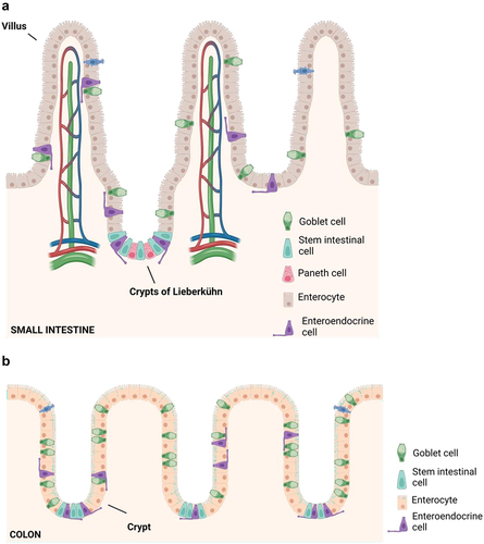
Figure 2. Gut mechanical barrier components. 1. Mucus layer is the outer mechanical barrier composed by mucus. 2. Epithelial cells: enterocytes, goblet cells, enteroendocrine cells, Paneth cells and microfold cells. 3. Tight junctions (TJs) are composed of proteins that control the paracellular pathway, as well as adherent junctions, desmosomes, and gap junctions. 4. Gut vascular barrier (GVB) constitutes the inner layer of defense, and it is composed by endothelial cells linked by TJs among others and different proteins that play a fundamental role regulating blood vessel permeability. Figure created with BioRender.com.
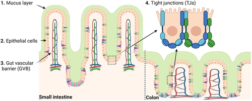
Figure 3. Photomicrograph of immunofluorescence (IF) preparation of colon stained with mucin-2 antibody. Nuclei are stained in blue. Staining was performed in 5 μm sections from paraffin embedded colon from a C57Bl/6J mouse 20 weeks age fed with chow diet. Scale = 100 μm.
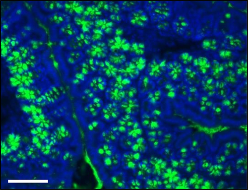
Figure 4. Schematic representation of TJ in gut. The paracellular pathway is controlled by a group of proteins known as junctional complexes that include, adherent junctions, gap junctions and tight junctions (composed by transmembrane proteins as claudins, occludin and junctional adhesion molecules (JAM)) which interact with the zona occludens (ZO) family of scaffolding proteins and the cytoskeletal actomyosin ring. Figure created with BioRender.com.
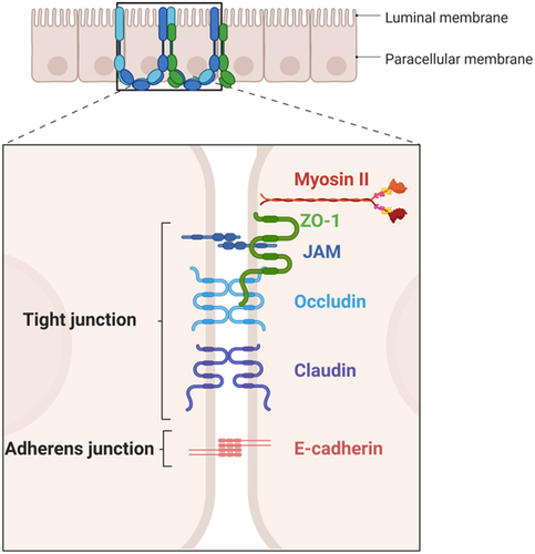
Figure 5. Photomicrograph of immunofluorescence (IF) preparation of colon stained with ZO-1. Nuclei are stained in blue. Staining was performed in 5 μm sections from paraffin embedded colon from a C57Bl/6J mouse 20 weeks age fed with chow diet. mounting. Scale = 100 μm.
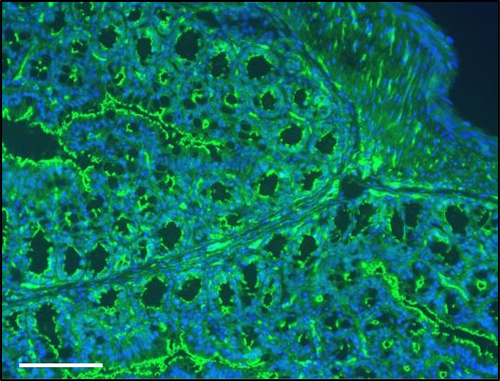
Figure 6. Representative photomicrograph of hematoxylin and eosin (H&E) staining in distal colon. Staining was performed in 5 μm sections from paraffin embedded colon from a C57Bl/6J mouse 20 weeks age fed with chow diet. GALT structure is marked with an arrow. Scale = 100 μm.
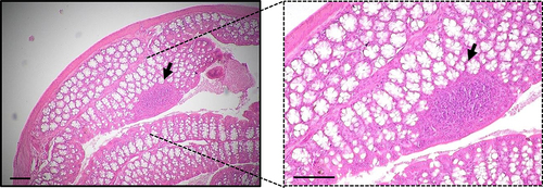
Figure 7. Immune barrier. Immune cells are mainly located in the gut mucose where secretory immunoglobulin a (SIgA) can be found. Gut barrier include macrophages, lymphocytes, paneth cells (more abundant in the small intestine) and microfold cells (M). Moreover, the gut-associated lymphoid tissue (GALT) is located in the lamina propria and can be classified into Peyer’s patches (PP) or isolated lymphoid follicles. Figure created with BioRender.com.
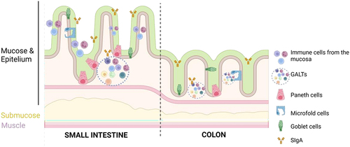
Figure 8. Enterohepatic BA cycle. Bile synthesis is performed in the liver by Cyp7a1 and Cyp27a11 enzymes. When synthesis is completed, BA are released in the bile canaliculi through the BSEP pump and stored in the gallbladder. In the ileum, bile salts are absorbed by ASBT and efluxed by OST-α/β to the circulation. Back to the liver they are uptaken by NTCP, a transporter located in the basolateral membrane of the hepatocytes. ABST, apical sodium-bile acid transporter; BSEP, bile salt export pump; FXR, farnesoid X receptor; NTCP, sodium taurocholate co-transporting polypeptide; OST, organic solute transporter.
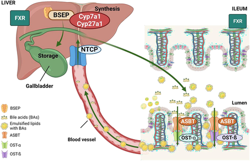
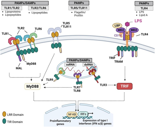
Figure 9. TLR types. TLR1, 2, 4, 5 and 6 bind to molecules associated with bacterial membranes. Concretely, TLR4, one of the most studied TLRs, binds to the LPS from the Gram-negative bacteria cell wall. TLR3, 7, 8 and 9 recognize viral, bacterial, or endogenous nucleic acids. Immune response initiation is mediated by TLR activation. DAMP, damage associated molecular patterns; LPS, lipopolysaccharide; PAMP, pathogen associated molecular patterns; TLR, toll-like receptor.
Breaking the barriers SUPPL.docx
Download MS Word (29.2 KB)Data availability statement
The authors confirm that the data supporting the findings of this study are available within the article.